 Indietro
Protein Structure and Function: Foundations of Biochemistry
Guida di studio - Note intelligenti
 Appunti personalizzati basati sui tuoi materiali, ampliati con definizioni chiave, esempi e contesto.
Protein Structure and Function
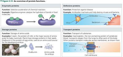
Main Functions of Proteins
Proteins are essential macromolecules that perform a vast array of functions in biological systems. Their diversity in structure allows them to participate in nearly every process within cells.
Enzymatic proteins: Catalyze biochemical reactions, increasing reaction rates. Example: Digestive enzymes hydrolyze food molecules.
Defensive proteins: Protect organisms from disease. Example: Antibodies neutralize pathogens.
Storage proteins: Store amino acids or other substances. Example: Ovalbumin in egg white stores amino acids for developing embryos.
Transport proteins: Move substances across cell membranes or throughout the body. Example: Hemoglobin transports oxygen in blood.
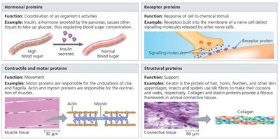
Hormonal proteins: Coordinate organismal activities. Example: Insulin regulates blood glucose levels.
Receptor proteins: Respond to chemical stimuli. Example: Receptors in nerve cells detect signaling molecules.
Contractile and motor proteins: Enable movement. Example: Actin and myosin in muscle contraction.
Structural proteins: Provide support and shape. Example: Collagen in connective tissues, keratin in hair and nails.
Chirality and Chemistry of Protein Building Blocks
Amino Acid Structure and Chirality
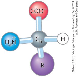
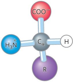
Amino acids are the monomeric units of proteins. Each amino acid (except glycine) contains a central (α) carbon atom bonded to four different groups: an amino group, a carboxyl group, a hydrogen atom, and a unique side chain (R group). This tetrahedral arrangement makes the α-carbon chiral, allowing for isomerism.
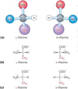
Chirality: Most amino acids are chiral (except glycine, whose R group is also hydrogen). Proteins are composed exclusively of L-amino acids.
Isomerism: L- and D- forms are non-superimposable mirror images (enantiomers). Only L-isomers are found in proteins.
Classification of Amino Acids
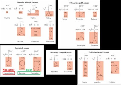
The 20 standard amino acids are classified based on the properties of their R groups:
Nonpolar (hydrophobic): 8 amino acids (e.g., Gly, Ala, Val, Leu, Ile, Met, Pro, Phe)
Polar (uncharged): 7 amino acids (e.g., Ser, Thr, Cys, Asn, Gln, Tyr, Trp)
Positively charged (basic): 3 amino acids (Lys, Arg, His)
Negatively charged (acidic): 2 amino acids (Asp, Glu)
Aromatic amino acids (Phe, Tyr, Trp) absorb UV light, which is useful for protein quantitation.
Peptide Bond Formation and Protein Primary Structure
Peptide Bond Formation
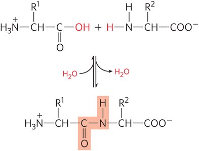
Proteins are formed by the condensation of amino acids, creating peptide bonds between the carboxyl group of one amino acid and the amino group of the next. This reaction releases water and occurs during translation on the ribosome.
Peptide bond: A covalent bond linking amino acids in a polypeptide chain.
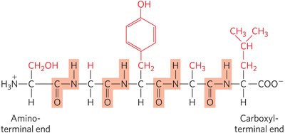
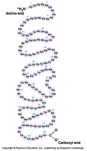
Directionality: Protein sequences are read from the amino (N) terminus to the carboxyl (C) terminus.
Primary Structure (1° Structure)
The primary structure of a protein is its unique sequence of amino acids, determined by the genetic code. This sequence dictates all higher levels of protein structure and function.
Importance: Determines folding, function, cellular location, activity, and evolutionary relationships.
Mutations: Changes in primary structure can lead to diseases (e.g., sickle cell anemia).
Diversity: With 20 amino acids, the number of possible sequences increases exponentially with chain length (e.g., for a tripeptide).
Protein Higher-Order Structure
Secondary Structure (2° Structure)
Secondary structure refers to local spatial arrangements of the polypeptide backbone, stabilized mainly by hydrogen bonds. The two most common types are the α-helix and β-sheet.
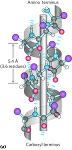
α-Helix: Right-handed coil stabilized by H-bonds between the C=O of residue n and the NH of residue n+4. Common in regulatory proteins, keratin, and hemoglobin. Ala and Leu favor helix formation; Gly and Pro disrupt it.
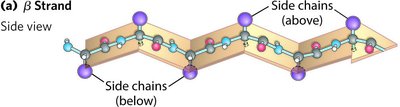
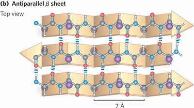
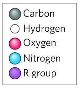
β-Sheet: Sheet-like structure formed by H-bonds between backbone atoms in different strands. Strands can be parallel or antiparallel. β-turns allow the chain to reverse direction.
Random coil: Irregular, non-repetitive regions of the polypeptide chain.
Tertiary Structure (3° Structure)
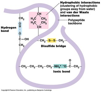
The tertiary structure is the overall three-dimensional arrangement of all atoms in a single polypeptide chain. It is stabilized by various interactions among side chains, including hydrophobic interactions, hydrogen bonds, ionic bonds, and disulfide bridges.
Hydrophobic effect: Nonpolar side chains cluster away from water, stabilizing the folded structure.
Disulfide bonds: Covalent bonds between cysteine residues add stability.
Electrostatic interactions: Salt bridges between charged side chains further stabilize the structure.
Quaternary Structure (4° Structure)
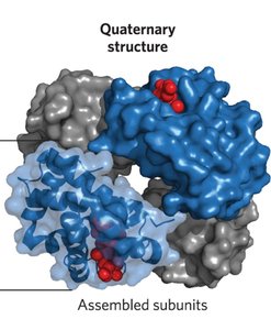
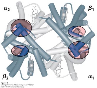
Quaternary structure arises when two or more polypeptide chains (subunits) assemble into a functional protein complex. Subunits may be identical (homomeric) or different (heteromeric).
Examples: Hemoglobin (α2β2 tetramer), antibodies, collagen, keratin.
Stabilization: Non-covalent interactions and sometimes covalent disulfide bonds.
Fibrous vs. Globular Proteins
Classification by Structure and Function
Proteins can be broadly classified based on their overall shape and function:
Fibrous proteins: Provide structural support, shape, and strength. Typically elongated and insoluble. Examples: Keratin (hair, nails), collagen (connective tissue), silk fibroin.
Globular proteins: Compact, soluble, and functionally diverse. Examples: Enzymes, antibodies, hemoglobin.
Examples of Fibrous Proteins
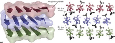
Silk fibroin: Composed of antiparallel β-sheets, rich in Ala and Gly, stabilized by close packing and hydrogen bonding.
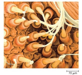
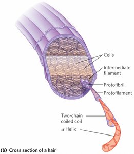
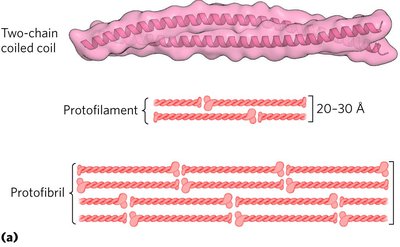
α-Keratin: Found in hair and nails, stabilized by disulfide bonds. Structure involves coiled coils and higher-order assemblies.
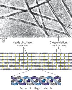
Collagen: Major component of connective tissue. Each chain forms a left-handed helix; three chains intertwine into a right-handed triple helix, providing high tensile strength.
Factors Affecting Protein Functionality
Protein function depends on correct folding and higher-order structure. Several factors can influence protein functionality:
Folding: May occur spontaneously or require assistance from chaperones.
Post-translational modifications: Chemical changes after synthesis can affect activity and localization.
Localization: Proper cellular compartmentalization is essential for function.
Conformational changes: Dynamic shifts in structure can regulate activity.
Denaturation: Loss of structure (and function) due to environmental changes.
Misfolding and degradation: Can lead to loss of function or disease.
