 Indietro
Protein Structure: From Peptide Bonds to Quaternary Organization
Guida di studio - Note intelligenti
 Appunti personalizzati basati sui tuoi materiali, ampliati con definizioni chiave, esempi e contesto.
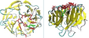
3-D Structure of Proteins
Overview of Protein Structure
Proteins are complex polymers of amino acids that fold into specific three-dimensional structures, which are essential for their biological functions. The organization of protein structure is hierarchical, ranging from the sequence of amino acids (primary structure) to the arrangement of multiple polypeptide chains (quaternary structure).
Primary structure: Linear sequence of amino acids linked by peptide bonds.
Secondary structure: Local folding patterns such as α-helices and β-sheets stabilized by hydrogen bonds.
Tertiary structure: Overall 3D folding of a single polypeptide chain.
Quaternary structure: Assembly of multiple polypeptide subunits.
Peptide Bond Formation and Properties
Condensation Reaction and Peptide Bond
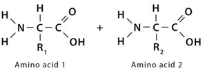
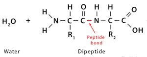
A peptide bond forms through a condensation (dehydration) reaction between the carboxyl group of one amino acid and the amino group of another, releasing water.
Reaction: Carboxyl group (–COOH) + Amino group (–NH2) → Peptide bond (–CO–NH–) + H2O
Result: Formation of a dipeptide and water.
Characteristics of the Peptide Bond
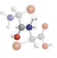
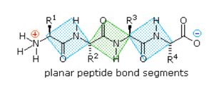
The peptide bond exhibits partial double-bond character due to resonance, making it planar and restricting rotation around the C–N bond.
Planarity: The peptide bond is rigid and planar, stabilizing the protein backbone.
Restricted rotation: Rotation is limited around the C–N bond, but possible around adjacent bonds (phi and psi angles).
Resonance: Delocalization of electrons between the carbonyl oxygen and the amide nitrogen.
Backbone Rotational Angles: Phi (φ), Psi (ψ), and Omega (ω)
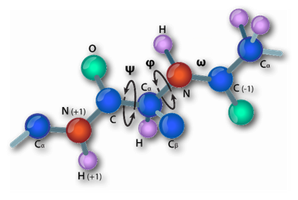
The flexibility of the polypeptide backbone is defined by three dihedral angles:
Phi (φ): Rotation around the N–Cα bond.
Psi (ψ): Rotation around the Cα–C bond.
Omega (ω): Rotation around the peptide bond (usually fixed at 180° due to planarity).
Secondary Structure: Alpha Helices
Alpha Helix Structure and Properties
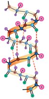
The α-helix is a common secondary structure where the polypeptide backbone coils into a right-handed spiral stabilized by hydrogen bonds.
Right-handed coil: Most naturally occurring α-helices are right-handed.
3.6 amino acids per turn and a pitch (vertical rise per turn) of 5.4 Å.
Hydrogen bonding: Between the C=O of residue i and the N–H of residue i+4.
Side chains: R-groups project outward, minimizing steric clashes.
Helical Variants: 310 and π-Helices
Variants of the α-helix include the 310-helix and π-helix, which differ in the number of residues per turn and hydrogen bonding patterns.
310-helix: 3 residues per turn, i→i+3 hydrogen bonding, 10 atoms in the H-bonded loop.
π-helix: 4.4 residues per turn, i→i+5 hydrogen bonding, 16 atoms in the H-bonded loop (rare).
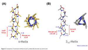
Helical Wheel Projection
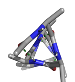
A helical wheel projection visualizes the spatial arrangement of side chains around the helix axis, useful for identifying amphipathic helices. 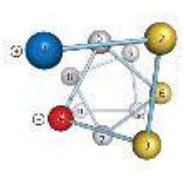
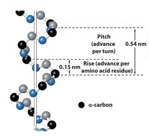
Pitch and Rise of an α-Helix
Pitch: Distance along the helix axis for one full turn (5.4 Å).
Rise: Vertical distance between adjacent α-carbons (1.5 Å per residue).
Peptide Bond Dipole
The peptide bond has a permanent dipole moment due to its resonance structure, contributing to a macroscopic dipole in α-helices that influences protein folding and function. 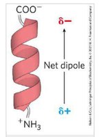
Secondary Structure: Beta Strands and Beta Sheets
Beta Strands and Beta Sheets
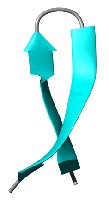
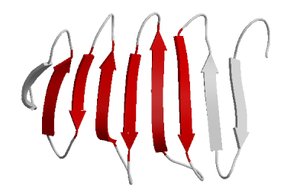
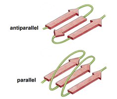
β-strands are extended polypeptide chains that align side-by-side to form β-sheets, stabilized by hydrogen bonds between backbone atoms.
Parallel β-sheets: Strands run in the same direction; hydrogen bonds are angled and slightly weaker.
Antiparallel β-sheets: Strands run in opposite directions; hydrogen bonds are linear and stronger.
Random Coils, Turns, and Loops
Random coils are flexible, disordered regions without regular secondary structure. Turns and loops connect secondary structure elements and often reverse the chain direction.
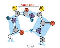
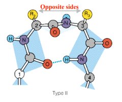
Turns (β-turns): Four amino acids forming a tight turn, stabilized by a hydrogen bond between the C=O of residue i and N–H of residue i+3.
Type I and II β-turns: Differ in the orientation of the peptide bond; Type II often contains glycine at position 3.
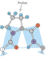
Gamma turns: Three amino acid turns, often involving proline.
Tertiary Structure
Stabilizing Interactions in Tertiary Structure
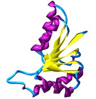
The tertiary structure is the overall 3D folding of a single polypeptide, stabilized by various interactions:
Hydrophobic interactions: Nonpolar side chains cluster away from water.
Hydrogen bonds: Between polar side chains and backbone atoms.
Ionic interactions: Between charged side chains.
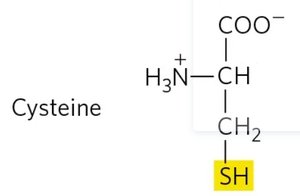
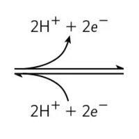
Disulfide bonds: Covalent bonds between cysteine residues.
Disulfide Bonds
Disulfide bonds are covalent links between the sulfur atoms of two cysteine residues, forming cystine. They stabilize protein structure, especially in extracellular proteins. 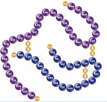
Hydrophobic and Aromatic Interactions
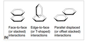
Hydrophobic side chains interact to minimize exposure to water, while aromatic rings can stabilize structure through π–π interactions (face-to-face, edge-to-face, parallel-displaced). 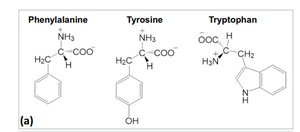
Quaternary Structure
Organization of Multiple Polypeptide Chains
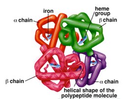
Quaternary structure refers to the arrangement of multiple polypeptide subunits in a functional protein complex. Interactions are similar to those stabilizing tertiary structure.
Examples: Hemoglobin (α2β2 tetramer), DNA polymerase, antibodies, ribosomes, ion channels.
Key Points
Proteins are polymers of amino acids with complex hierarchical structures.
Peptide bonds are planar and restrict backbone rotation, influencing protein folding.
Secondary structures (α-helix, β-sheet) are stabilized by hydrogen bonds.
Tertiary and quaternary structures are stabilized by hydrophobic, ionic, hydrogen bonding, and covalent (disulfide) interactions.
Structure determines protein function; denaturation disrupts this relationship.
