 Indietro
The Three-Dimensional Structure of Proteins
Guida di studio - Note intelligenti
 Appunti personalizzati basati sui tuoi materiali, ampliati con definizioni chiave, esempi e contesto.
Chapter 6: The Three-Dimensional Structure of Proteins
Introduction
The three-dimensional structure of proteins is fundamental to their biological function. This chapter explores the hierarchical organization of protein structure, the principles governing secondary structure formation, the role of amino acid properties, and the mechanisms of protein folding and misfolding.
Levels of Protein Structure
Primary Structure
The primary structure of a protein is its unique sequence of amino acids, linked by peptide bonds. This sequence determines all higher levels of structure and ultimately the protein's function.
Peptide bond: The covalent bond between the carboxyl group of one amino acid and the amino group of the next.
Directionality: Proteins are synthesized from the N-terminus to the C-terminus.
Secondary Structure
Secondary structures are local, regularly repeating structures stabilized by hydrogen bonds between backbone atoms. The main types are the α-helix, β-sheet, and 310-helix.
α-helix: Right-handed coil with 3.6 residues per turn, stabilized by hydrogen bonds between the carbonyl oxygen of residue i and the amide hydrogen of residue i+4.
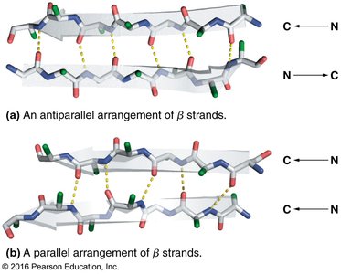
β-sheet: Extended strands connected laterally by hydrogen bonds, can be parallel or antiparallel.
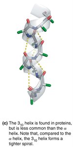
310-helix: Tighter helix with 3 residues per turn and hydrogen bonds between residues i and i+3.
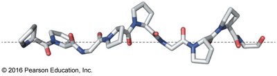
Polypeptide II helix: Left-handed helix, common in collagen, not stabilized by hydrogen bonds.
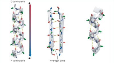
Hydrogen Bonding in Secondary Structures
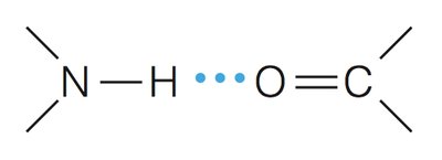
Hydrogen bonds are the primary stabilizing force in both α-helices and β-sheets.
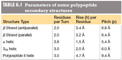
Parameters of Secondary Structures
Structure Type | Residues per Turn | Rise (h) per Residue | Pitch (p) |
|---|---|---|---|
β Strand (antiparallel) | 2.0 | 3.4 Å | 6.8 Å |
β Strand (parallel) | 2.0 | 3.2 Å | 6.4 Å |
α helix | 3.6 | 1.5 Å | 5.4 Å |
310 helix | 3.0 | 2.0 Å | 6.0 Å |
Polypeptide II helix | 3.0 | 4.7 Å | 9.4 Å |
Tertiary Structure
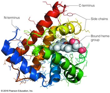
The tertiary structure is the overall three-dimensional arrangement of all atoms in a single polypeptide chain, including the spatial arrangement of secondary structure elements and side chains.
Stabilized by hydrophobic interactions, hydrogen bonds, ionic interactions, van der Waals forces, and sometimes disulfide bonds.
Domains: Independently folded regions with specific functions.
Quaternary Structure
Quaternary structure refers to the spatial arrangement of multiple polypeptide chains (subunits) in a multisubunit complex.
Subunits may be identical (homotypic) or different (heterotypic).
Examples: Hemoglobin (α2β2 tetramer), transthyretin dimer.
Principles Governing Protein Structure
Bond Angles and Planarity
Linus Pauling established rules for secondary structure formation:
Bond angles and lengths are similar to those in free amino acids.
No atoms approach closer than their van der Waals radii.
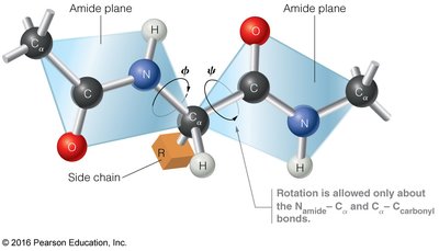
The amide group is planar due to partial double-bond character of the peptide bond.
Noncovalent bonds, especially hydrogen bonds, stabilize the structure.
Backbone Dihedral Angles (ϕ and ψ)
Rotation in the polypeptide backbone is restricted to the Namide-Cα (ϕ) and Cα-Ccarbonyl (ψ) bonds. The peptide bond itself is not rotatable.
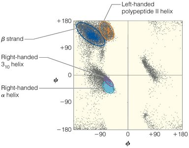
Ramachandran Plot
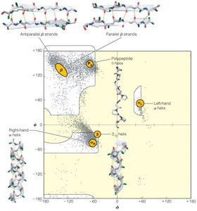
The Ramachandran plot displays the sterically allowed combinations of ϕ and ψ angles for amino acid residues in a polypeptide. Most conformations are forbidden due to steric clashes.
Glycine, with a small side chain, has more allowed conformations.
Proline, with a cyclic side chain, is highly restricted.
Secondary Structure Features
α-Helix
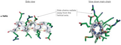
3.6 residues per turn, 5.4 Å pitch.
Side chains radiate outward from the helix axis.
Hydrogen bonds are nearly parallel to the helix axis.
Often amphiphilic, with distinct hydrophilic and hydrophobic faces.
β-Sheet
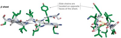
Composed of β-strands connected laterally by hydrogen bonds.
Strands can be parallel or antiparallel.
Side chains alternate above and below the plane of the sheet.
310-Helix and Polypeptide II Helix
310-helix: 3 residues per turn, tighter than α-helix, less common.
Polypeptide II helix: Left-handed, not stabilized by hydrogen bonds, prevalent in collagen.
Protein Folding and Stability
Anfinsen’s Experiment
Christian Anfinsen demonstrated that the information required for protein folding is contained in the amino acid sequence. Denatured ribonuclease A can refold into its native structure, supporting the "thermodynamic hypothesis" of protein folding.
Thermodynamic Factors in Folding
Favorable enthalpy: Intramolecular interactions (hydrogen bonds, ionic bonds, van der Waals forces).
Unfavorable entropy: Loss of conformational freedom upon folding.
Favorable solvent entropy: Hydrophobic effect—burying nonpolar side chains releases ordered water molecules.
Disulfide Bonds
Disulfide bonds between cysteine residues stabilize protein structure, especially in extracellular proteins, by reducing the number of possible unfolded conformations.
Protein Folding Pathways
Proteins do not sample all possible conformations (Levinthal’s paradox). Folding proceeds through intermediates, such as the "molten globule," and is often assisted by molecular chaperones.
Chaperones: Proteins that assist in proper folding and prevent aggregation.
Chaperonins: Large protein complexes (e.g., GroEL/ES) that provide an isolated environment for folding.
Protein Misfolding and Disease
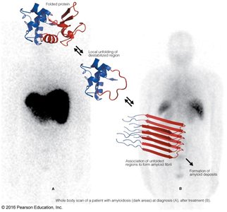
Misfolded proteins can aggregate to form amyloid fibrils, associated with diseases such as amyloidosis and prion diseases. Amyloid formation involves the association of unfolded regions into ordered fibrils.
Classification of Protein Structure
Fibrous vs. Globular Proteins
Fibrous proteins: Long fibers or sheets, mechanically strong, insoluble in water, structural roles (e.g., keratin, collagen).
Globular proteins: Compact, spherical, soluble, diverse functions (e.g., enzymes, myoglobin).
Amino Acid Distribution
In globular proteins, hydrophobic residues are typically buried in the interior, while hydrophilic residues are exposed to the solvent. This distribution drives the folding process and stabilizes the native structure.
Prediction of Protein Structure
Secondary Structure Prediction
Empirical methods use amino acid propensities for helix or sheet formation.
Amphiphilic helices and strands show repeating patterns of side chain polarity.
Tertiary Structure Prediction
More challenging due to long-range interactions.
Current computational methods are about 60% accurate.
Summary Table: Ranges of Allowed ϕ and ψ Angles
Structure Type | ϕ | ψ |
|---|---|---|
β strand | −150° to −100° | +120° to +160° |
α helix | −70° to −60° | −50° to −40° |
310 helix | −70° to −60° | −30° to −10° |
Polypeptide II helix | −80° to −60° | +130° to +160° |
Key Equations
Beer-Lambert Law:
Hydrogen bond:
Conclusion
Understanding the three-dimensional structure of proteins is essential for grasping their function, mechanisms of folding, and the consequences of misfolding. The interplay of chemical forces, sequence, and structure underlies the remarkable diversity and specificity of protein function in biological systems.
