 Indietro
Three-Dimensional Structure of Proteins: Secondary, Tertiary, and Quaternary Organization
Guida di studio - Note intelligenti
 Appunti personalizzati basati sui tuoi materiali, ampliati con definizioni chiave, esempi e contesto.
Three-Dimensional Structure of Proteins
Overview of Protein Functions
Proteins are essential biomolecules that perform a wide variety of functions in living organisms. Their function is determined by their three-dimensional structure, which is stabilized by various interactions.
Structural Proteins: Provide mechanical support (e.g., collagen, elastin).
Enzymes: Biological catalysts for cellular reactions.
Transport and Storage: Carry small biomolecules (e.g., hemoglobin, ferritin).
Muscle Contraction and Mobility: Actin and myosin are key components.
Immune Proteins: Defensive proteins such as antibodies.
Regulatory and Receptor Proteins: Mediate cellular activity and signaling (e.g., hormones, G-proteins).
Protein function depends on selective interactions with other molecules, primarily through non-covalent interactions (ionic, hydrogen bonds, van der Waals, hydrophobic effect).

Levels of Protein Structure
Proteins are organized into four structural levels, each contributing to their overall shape and function.
Primary Structure: Linear sequence of amino acids held by covalent peptide bonds.
Secondary Structure: Local 3D arrangement of the backbone, stabilized by hydrogen bonds (e.g., α-helix, β-sheet).
Tertiary Structure: Overall spatial arrangement of all atoms, including side chains and prosthetic groups.
Quaternary Structure: Assembly of multiple polypeptide chains into complexes.
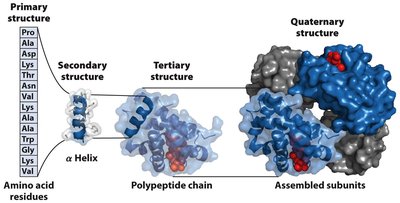
Forces Stabilizing Protein Structure
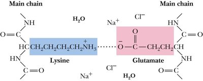
Secondary, tertiary, and quaternary structures are stabilized by weak forces:
Hydrogen bonds: Formed wherever possible.
Hydrophobic interactions: Drive protein folding.
Ionic interactions: Usually occur on the protein surface.
Van der Waals interactions: Ubiquitous throughout the protein.
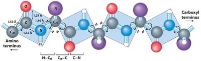
Origin of 3D Structure: Rotation Around Bonds
Peptide Bond Rigidity and Rotation
The peptide bond is planar and rigid due to partial double bond character, preventing rotation. Free rotation is allowed around the α-carbon (Cα) bonds, described by two dihedral angles:
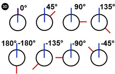
ϕ (phi): Angle around the α-carbon—amide nitrogen bond.
ψ (psi): Angle around the α-carbon—carbonyl carbon bond.




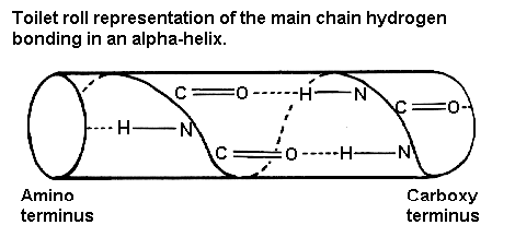
Secondary Structure
α-Helix
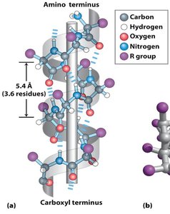
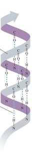
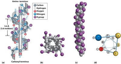
The α-helix is a rod-like structure stabilized by hydrogen bonds between the carbonyl oxygen of one amino acid and the amide hydrogen four residues later (i+4). It is right-handed, with 3.6 residues per turn and a pitch of 5.4 Å.
Side chains: Point outward, perpendicular to the helix axis.
Stabilizing factors: Hydrogen bonds parallel to the helix axis.
Helix breakers: Proline (impossible rotation), Glycine (too mobile).
β-Sheet
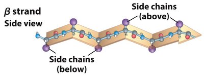
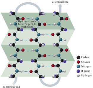
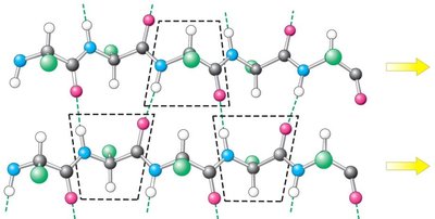
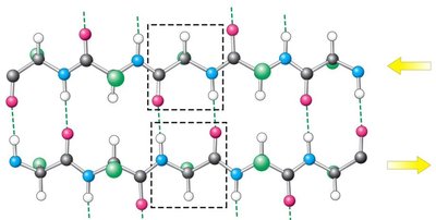
β-sheets consist of β-strands held together by hydrogen bonds between backbone amide and carbonyl groups. Side chains alternate above and below the sheet.
Parallel β-sheets: Strands run in the same direction; hydrogen bonds are bent and weaker.
Antiparallel β-sheets: Strands run in opposite directions; hydrogen bonds are linear and stronger.
β-Turns (Loops)
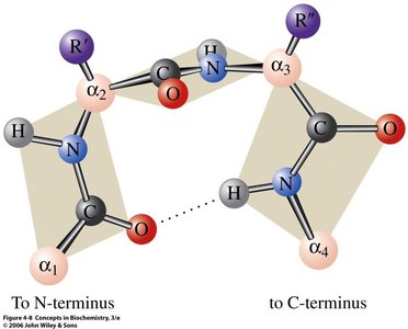
β-turns are small regions that allow the peptide chain to reverse direction, often stabilized by hydrogen bonds. Proline and glycine are common in β-turns due to their structural properties.
Steric Interactions and Allowed Angles
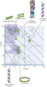
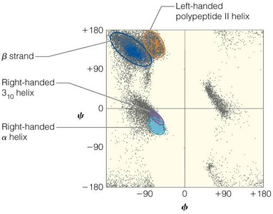
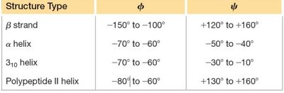
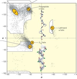
Steric clashes restrict the possible values of ϕ and ψ angles, limiting the conformations proteins can adopt. The Ramachandran plot shows the sterically allowed regions for these angles.
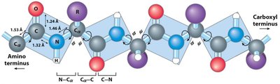
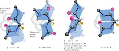
Table: Idealized ϕ and ψ Angles for Common Secondary Structures
Structure | ϕ | ψ |
|---|---|---|
α Helix | –57˚ | –47˚ |
β Conformation (Antiparallel) | –139˚ | +135˚ |
β Conformation (Parallel) | –119˚ | +113˚ |
Collagen triple helix | –51˚ | +153˚ |
β Turn type I (i+1a) | –60˚ | –30˚ |
β Turn type I (i+2a) | –90˚ | 0˚ |
β Turn type II (i+1) | –60˚ | +120˚ |
β Turn type II (i+2) | +80˚ | 0˚ |
Ramachandran Plot
The Ramachandran plot is a graphical representation of the allowed regions for ϕ and ψ angles in proteins. It helps predict secondary structure elements and reveals regions with unusual backbone structure.
Tertiary Structure
Forces Stabilizing Tertiary Structure
Tertiary structure is the overall spatial arrangement of all atoms in a protein, stabilized by interactions among side chains:
Hydrophobic interactions: Nonpolar side chains cluster in the interior.
Ionic/electrostatic interactions: Charged side chains interact, often on the surface.
Hydrogen bonding: Among polar side chains.
Disulfide bridges: Covalent bonds between cysteine residues.
Metal ion coordination: Metal ions bind to negatively charged side chains.
Classification of Proteins by Tertiary Structure
Fibrous Proteins: Polypeptide chains organized parallel to a single axis, mechanically strong, insoluble, structural role (e.g., keratin, collagen, fibroin).
Globular Proteins: Diverse, flexible, more numerous, functional roles (e.g., enzymes, regulatory proteins).
α-Keratin and Coiled-Coil Structure
α-Keratin is a fibrous protein with repeating hydrophobic residues, forming a coiled-coil structure stabilized by disulfide bonds. This structure gives elasticity and strength to hair, wool, and nails.
Silk Fibroin and Collagen
Silk fibroin is composed of antiparallel β-sheets, rich in glycine and alanine, allowing close packing and strength. Collagen forms a triple helix, stabilized by hydroxyproline and hydroxylysine, essential for connective tissue integrity.
Globular Proteins
Globular proteins have a nonpolar interior and a hydrophilic exterior, often containing distinct domains with specific functions. Their surfaces are composed of loops and turns, enabling interactions with other molecules.
Quaternary Structure
Assembly of Subunits
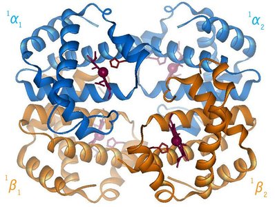
Quaternary structure involves the assembly of two or more polypeptide chains into functional complexes. Subunits interact through non-covalent interactions, and the association provides stability, functional catalytic sites, and cooperativity.
Homomultimers: Single type of monomer.
Heteromultimers: Different types of monomers.
Denaturation and Protein Folding
Denaturation
Denaturation is the loss of protein structure and function due to external stresses (heat, chemicals, pH changes). The primary structure remains intact, but secondary, tertiary, and quaternary structures are disrupted.
Denaturing agents: Heat, agitation, pH, β-mercaptoethanol, detergents, urea, guanidine HCl.
Protein Folding
Protein folding is a spontaneous process driven by the hydrophobic effect, hydrogen bonds, and van der Waals forces. The goal is to achieve the lowest energy state, with the information for folding encoded in the primary sequence.
Levinthal’s paradox: Folding cannot occur by random sampling; secondary structures form first, followed by hydrophobic collapse and long-range interactions.
Free-energy funnel model: Folding involves intermediate states, with the native structure at the lowest free-energy point.
Diseases Associated with Protein Structure Defects
Cystic Fibrosis
Cystic fibrosis is caused by mutations in the CFTR gene, affecting chloride ion transport and leading to thickened mucus and various symptoms. The most severe mutation is the deletion of phenylalanine at position 508, causing the protein to misfold and get stuck in the endoplasmic reticulum.
Symptoms: Breathing problems, respiratory infections, excessive mucus, salty skin, sterility in males.
Treatments: Symptomatic therapy, gene therapy.
Practice and Review
Practice Questions
Which peptide could form an amphiphilic α-helix?
Which of the following statements is FALSE?
Which of the following statements is correct?
Refer to textbook for end-of-chapter problems and answers.
