Indietro
Membrane Structure and Transport: General Biology Study Notes
Guida di studio - Note intelligenti
 Appunti personalizzati basati sui tuoi materiali, ampliati con definizioni chiave, esempi e contesto.
Membrane Structure & Transport
Phospholipids: Structure and Properties
Phospholipids are the fundamental building blocks of cellular membranes. Their unique structure allows them to form bilayers that separate the internal environment of the cell from the external environment.
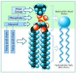
Structure: Phospholipids consist of a glycerol backbone, two fatty acid chains (hydrophobic tails), and a phosphate group (hydrophilic head).
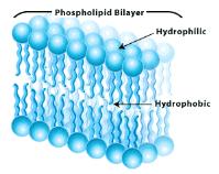
Amphipathic Nature: The molecule contains both hydrophilic (polar) and hydrophobic (nonpolar) regions, making it amphipathic.
Hydrophilic Head: Interacts with water due to polar covalent bonds and charged atoms.
Hydrophobic Tails: Consist of nonpolar hydrocarbon chains that avoid water.
Role: Phospholipids spontaneously form bilayers in aqueous environments, establishing boundaries for cells and organelles.
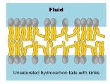
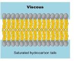
Saturated vs. Unsaturated: Saturated fatty acids have no double bonds, making tails straight and allowing tight packing. Unsaturated fatty acids have one or more double bonds, causing kinks and preventing tight packing.
Example: Cell membranes are composed primarily of phospholipid bilayers.
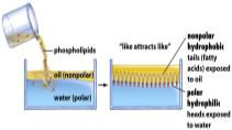
Amphipathic Lipids and Water
Amphipathic lipids interact with water and oil in distinct ways, leading to the formation of specialized structures.
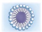
Micelles: Single-layered structures with hydrophobic tails inward and hydrophilic heads outward.
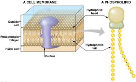
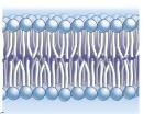
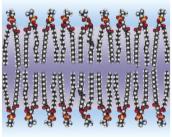
Lipid Bilayers: Two sheets of phospholipids with hydrophilic heads facing water and hydrophobic tails facing each other.
Spontaneous Formation: Bilayers form without energy input due to the amphipathic nature of phospholipids.
Example: Oil and water separation demonstrates how phospholipids orient themselves at interfaces.
Membrane Structure: Bilayers and Vesicles
Phospholipid bilayers are the basis for biological membranes, forming boundaries and compartments within cells.
Lipid Bilayer: Hydrophilic heads face outward, hydrophobic tails face inward.
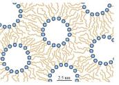
Vesicles: Small spheres surrounded by lipid bilayers, used for transport within cells.
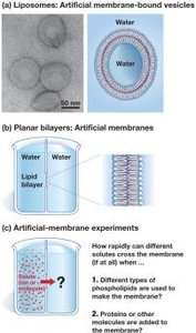
Artificial Membranes: Liposomes and planar bilayers can be created in the lab to study membrane properties.
Example: Laboratory experiments use liposomes to mimic cell membranes.
Membrane Permeability
Selective Permeability of Phospholipid Bilayers
Cell membranes are selectively permeable, allowing certain molecules to pass while restricting others.
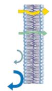
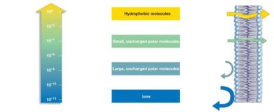
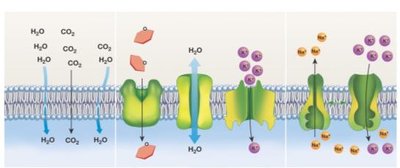
Small, Nonpolar Molecules: Move across membranes easily (e.g., O2, CO2).
Large or Charged Molecules: Cross slowly or not at all (e.g., glucose, ions).
Permeability Scale: Hydrophobic molecules have the highest permeability, ions the lowest.
Factors Affecting Permeability: Molecular composition (saturated vs. unsaturated fatty acids), presence of proteins, cholesterol, and temperature.
Unsaturated Fatty Acids: Increase permeability due to kinks preventing tight packing.
Saturated Fatty Acids: Decrease permeability due to tight packing.
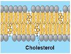
Cholesterol: Modifies membrane fluidity and permeability.


Temperature and Membrane Properties
Temperature influences membrane fluidity and permeability by affecting the motion of lipid molecules.
Phase Transition: At a specific melting temperature (Tm), membranes shift from a gel phase (tight packing) to a liquid crystalline phase (loose packing).
Increased Kinetic Energy: Above Tm, hydrocarbon chains become more flexible, increasing surface area and permeability.
Regulation: Cells adjust lipid, protein, and cholesterol content to regulate permeability.
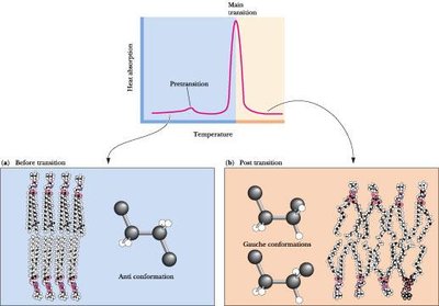
Fluidity vs. Permeability
Membrane fluidity refers to the movement of molecules within the bilayer, which is closely linked to permeability.
Direct Relationship: Increased fluidity leads to increased permeability.
Factors: Elevated temperature, unsaturated fatty acids, and cholesterol increase fluidity.
Example: Membrane components are in constant motion, similar to objects floating on water.
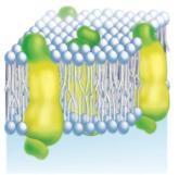
Fluid Mosaic Model of Membrane Structure
Fluid Mosaic Model
The fluid mosaic model describes the dynamic nature of cell membranes, composed of a mosaic of lipids and proteins.
Integral Proteins: Span the membrane, with segments facing both interior and exterior.
Peripheral Proteins: Located on one side of the membrane.
Dynamic Mosaic: Lipids and proteins move freely within the bilayer.
Experimental Evidence: Cell fusion experiments demonstrate protein mobility, supporting the fluid mosaic model.

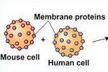
Membrane Transport Mechanisms
Diffusion and Equilibrium
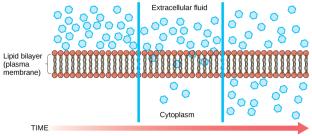
Diffusion is the spontaneous movement of molecules from regions of high concentration to low concentration, establishing equilibrium across membranes.
Concentration Gradient: Drives net movement of solutes.
Electrochemical Gradient: Ions create both concentration and electrical gradients.
Example: Small, nonpolar molecules move across membranes by diffusion.

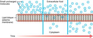
Osmosis
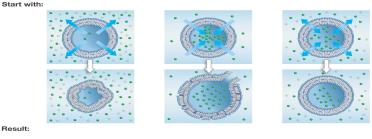
Osmosis is the diffusion of water across a selectively permeable membrane in response to solute concentration differences.
Hypertonic Solution: Higher solute concentration outside; water leaves cell, cell shrinks.
Hypotonic Solution: Lower solute concentration outside; water enters cell, cell swells or bursts.
Isotonic Solution: Equal solute concentration; cell size remains unchanged.
Biological Relevance: Osmosis can shrink or burst cells, affecting their function.
Facilitated Diffusion
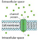
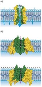
Facilitated diffusion is passive transport of molecules across membranes via specific proteins, allowing movement of substances that cannot cross the bilayer directly.
Channel Proteins: Form pores for specific ions or molecules.
Carrier Proteins: Undergo conformational changes to transport substances.
Electrochemical Gradients: Drive ion movement through channels.
Example: Aquaporins facilitate water transport; potassium channels allow K+ ions to pass.
Active Transport
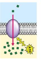
Active transport moves molecules or ions against their concentration or electrochemical gradients, requiring energy input (ATP) and specialized proteins called pumps.
ATP Hydrolysis: Provides energy for transport.
Pumps: Transmembrane proteins that move substances across membranes.
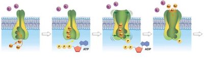
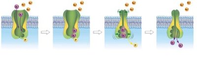
Sodium-Potassium Pump (Na+/K+-ATPase): Transports Na+ out and K+ into the cell, maintaining gradients essential for cell function.
Example: The Na+/K+ pump operates in a cycle of binding, phosphorylation, conformational change, and release.
Bulk Transport: Exocytosis and Endocytosis
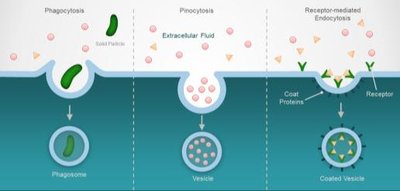
Bulk transport mechanisms move large quantities of materials into or out of cells via vesicles, requiring energy (ATP).
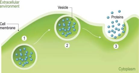
Exocytosis: Export of molecules via vesicles that fuse with the cell membrane.
Endocytosis: Import of materials via vesicles; includes phagocytosis (cells), pinocytosis (fluids), and receptor-mediated endocytosis.
Example: Cells use endocytosis to take up nutrients and exocytosis to secrete proteins.