 Indietro
Proteins and Nucleic Acids: Structure and Function
Guida di studio - Note intelligenti
 Appunti personalizzati basati sui tuoi materiali, ampliati con definizioni chiave, esempi e contesto.
Proteins: Structure and Function
Overview of Protein Diversity
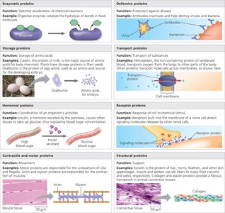
Proteins are essential macromolecules that perform a vast array of functions in living organisms. Each protein has a unique three-dimensional structure, which determines its specific function. Humans possess tens of thousands of different proteins, each constructed from the same set of 20 amino acids.
Key Point 1: Proteins are responsible for nearly every dynamic function in living beings, including catalysis, defense, transport, signaling, movement, and structure.
Key Point 2: The diversity of protein structure leads to a wide range of functions.
Example: Enzymes, antibodies, transport proteins, and structural proteins each have distinct roles in the cell.
Amino Acids: The Building Blocks of Proteins
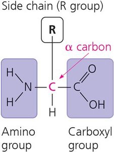
Amino acids are organic molecules possessing both a carboxyl and an amino group. They are the monomers of polypeptides, and all amino acids share a common structure centered around an asymmetric carbon atom (alpha carbon) with four different partners: amino group, carboxyl group, hydrogen atom, and an R group (side chain).
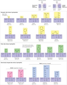
Key Point 1: The R group (side chain) varies among amino acids and determines their unique characteristics.
Key Point 2: The physical and chemical properties of the side chain affect the functional role of the amino acid in a polypeptide.
Example: Glycine has a simple hydrogen R group, while glutamic acid has a complex, charged side chain.
Polypeptides and Peptide Bonds
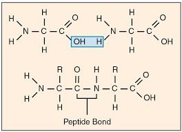
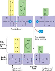
Polypeptides are polymers of amino acids linked together by peptide bonds, which are covalent bonds formed by dehydration reactions between the carboxyl group of one amino acid and the amino group of another. Each polypeptide has a unique linear sequence of amino acids, and the sequence and nature of the side chains determine how the polypeptide folds and its final shape.
Key Point 1: Peptide bonds are formed through dehydration reactions.
Key Point 2: The sequence of amino acids (primary structure) is critical for protein function.
Example: A change in the sequence can lead to diseases such as sickle cell anemia.
Levels of Protein Structure
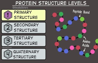
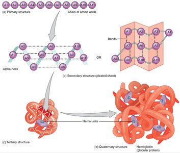
Proteins exhibit four levels of structure: primary, secondary, tertiary, and quaternary. The primary structure is the linear sequence of amino acids. Secondary structure refers to regions of repetitive coiling or folding (alpha helices and beta sheets) stabilized by hydrogen bonds. Tertiary structure is the overall three-dimensional shape stabilized by interactions between side chains. Quaternary structure arises when a protein consists of two or more polypeptide chains.
Key Point 1: Each level of structure is essential for the protein's function.
Key Point 2: The function of a protein depends on its ability to recognize and bind to other molecules.
Example: Hemoglobin is a quaternary structure protein composed of four polypeptide chains.
Protein Structure and Disease
Even a slight change in the primary structure can affect a protein’s shape and ability to function. Sickle cell disease is caused by a single nucleotide change in the gene encoding hemoglobin, resulting in abnormal aggregation and altered red blood cell shape.
Key Point 1: Protein structure is sensitive to changes in amino acid sequence.
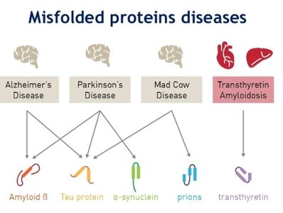
Key Point 2: Misfolded proteins are associated with diseases such as cystic fibrosis, Alzheimer’s, Parkinson’s, and mad cow disease.
Example: Sickle cell crises occur when misshapen cells clog blood vessels.
Denaturation and Protein Folding
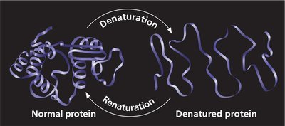
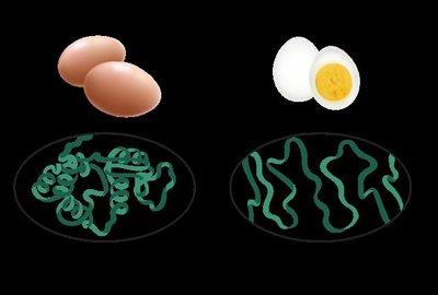
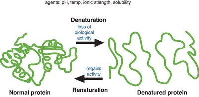
Protein structure depends on environmental conditions such as pH, salt concentration, and temperature. Denaturation is the process in which a protein loses its native shape due to disruption of weak chemical bonds and interactions, rendering it biologically inactive. Excessive heat, chemicals, or transfer to a nonpolar solvent can cause denaturation.
Key Point 1: Denatured proteins lose their biological activity.
Key Point 2: Some proteins can regain their structure through renaturation.
Example: Cooking an egg denatures its proteins, causing them to solidify.
Protein Folding and Structural Determination
Misfolding of polypeptides is a serious problem in cells. X-ray crystallography is the most common method used to determine the three-dimensional structure of a protein, relying on the diffraction of X-rays by the atoms in a crystallized molecule.
Key Point 1: Proper folding is essential for protein function.
Key Point 2: X-ray crystallography provides detailed structural information.
Example: Structural determination helps understand protein function and disease mechanisms.
Nucleic Acids: Structure and Function
Overview of Nucleic Acids
Nucleic acids are polymers made of monomers called nucleotides. The amino acid sequence of a polypeptide is programmed by a gene, which consists of DNA. There are two types of nucleic acids: deoxyribonucleic acid (DNA) and ribonucleic acid (RNA).
Key Point 1: DNA provides directions for its own replication and directs RNA synthesis.
Key Point 2: RNA is involved in protein synthesis and gene regulation.
Example: DNA determines the inherited structure of a cell’s proteins.
Components of Nucleic Acids
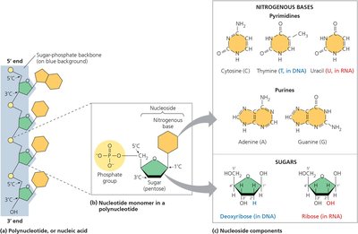
Polynucleotides are polymers consisting of many nucleotide monomers in a chain. Each nucleotide consists of a five-carbon sugar (pentose), a nitrogen-containing base, and one to three phosphate groups. The portion of a nucleotide without any phosphate groups is called a nucleoside.
Key Point 1: There are two families of nitrogenous bases: pyrimidines (C, T, U) and purines (A, G).
Key Point 2: DNA contains deoxyribose, while RNA contains ribose.
Example: Thymine is found only in DNA, uracil only in RNA.
Nucleotide Polymers and Directionality
Nucleotides are linked into polynucleotides by phosphodiester linkages, forming a sugar-phosphate backbone. The two free ends of the polymer are different: one has a phosphate attached to a 5’ carbon, the other a hydroxyl group on a 3’ carbon. Polynucleotides have directionality from 5’ to 3’.
Key Point 1: The sequence of bases along a DNA polymer is unique for each gene.
Key Point 2: The linear order of bases specifies the amino acid sequence of a protein.
Example: The sequence 5′-AGGTAACTT-3′ encodes specific genetic information.
Structures of DNA and RNA Molecules
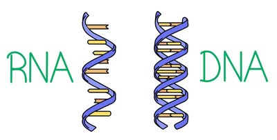
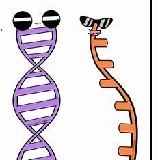
Native DNA forms a double helix, with two antiparallel polynucleotide strands wound around an axis. The two strands are held together by hydrogen bonds between paired bases: adenine pairs with thymine, guanine pairs with cytosine. RNA molecules exist as single strands, but complementary base pairing can occur within or between RNA molecules, allowing them to take on specific three-dimensional shapes.
Key Point 1: DNA double helix is stabilized by base pairing.
Key Point 2: RNA base pairing allows for functional three-dimensional shapes.
Example: In RNA, adenine pairs with uracil; thymine is not present.
Genetic Information Flow
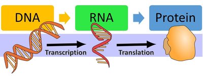
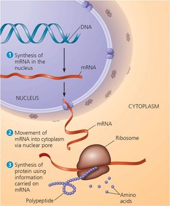
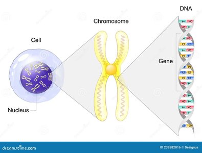
DNA is the genetic material inherited from parents. Each chromosome contains one long DNA molecule carrying hundreds of genes. When a cell divides, its DNA is copied and passed to the next generation. Messenger RNA (mRNA) is synthesized from DNA and directs protein synthesis at ribosomes in the cytoplasm.
Key Point 1: DNA encodes genetic information, but proteins implement genetic programs.
Key Point 2: mRNA conveys genetic instructions from the nucleus to the cytoplasm.
Example: The central dogma: DNA → RNA → Protein.
Summary Table: Protein and Nucleic Acid Comparison
Feature | Proteins | Nucleic Acids |
|---|---|---|
Monomer | Amino acid | Nucleotide |
Polymer | Polypeptide | Polynucleotide |
Bond Type | Peptide bond | Phosphodiester bond |
Function | Catalysis, structure, transport, signaling | Genetic information storage and transfer |
Structure | Primary, secondary, tertiary, quaternary | Double helix (DNA), single strand (RNA) |
