 Indietro
A Preview of Cell Biology: Foundations, Microscopy, and the Cell Theory
Guida di studio - Note intelligenti
 Appunti personalizzati basati sui tuoi materiali, ampliati con definizioni chiave, esempi e contesto.
A Preview of Cell Biology
Introduction to Cell Biology
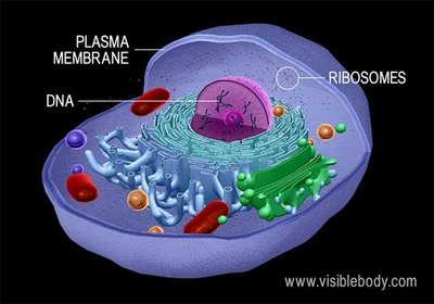
Cell biology is the study of cells, the fundamental units of life. Cells possess the ability to grow, reproduce, and differentiate into specialized types. The integration of cytology, genetics, and biochemistry has transformed cell biology into a dynamic and foundational discipline in modern biology.
Cell: The basic structural and functional unit of all living organisms.
Specialization: Cells can differentiate to perform unique functions within multicellular organisms.
Modern Cell Biology: Emerged from the convergence of cytology (cell structure), genetics (heredity and information flow), and biochemistry (cellular chemistry).
The Cell Theory: Historical Foundations
Early Observations and the Birth of Cell Theory
The development of cell theory was driven by advances in microscopy and careful observation. Early scientists laid the groundwork for our understanding of cells as the basic units of life.
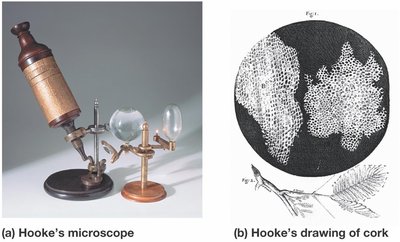
Robert Hooke (1665): First observed and named 'cells' while examining cork tissue under a microscope. The term 'cell' comes from the Latin cellula, meaning 'little room'.

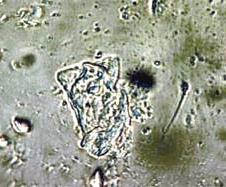
Antonie van Leeuwenhoek (1673): Improved lens technology, achieving up to 300x magnification. He was the first to observe living single cells, including bacteria and protozoa, which he called 'animalcules'.

Technological Limitations: Early microscopes had low resolution, and cell biology was initially descriptive rather than explanatory.
Development of Compound Microscopes and Discovery of the Nucleus
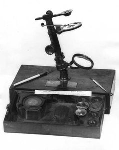
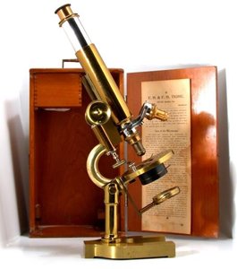
By the 1830s, compound microscopes with two lenses improved magnification and resolution, allowing scientists to observe structures as small as 1 micrometer.
Robert Brown: Identified the nucleus within plant cells.
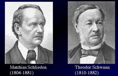
Matthias Schleiden and Theodor Schwann: Concluded that all plant and animal tissues are composed of cells.
The Cell Theory
Formulated in 1839 by Theodor Schwann and later expanded by Rudolf Virchow, the cell theory is a cornerstone of biology.
1. All organisms consist of one or more cells.
2. The cell is the basic unit of structure for all organisms.
3. All cells arise only from preexisting cells (Virchow, 1855).

The Emergence of Modern Cell Biology
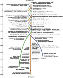
Three Strands of Inquiry
Modern cell biology integrates three major strands of biological inquiry:
Cytology: Focuses on cellular structure using optical techniques.
Biochemistry: Examines cellular structure and function at the molecular level.
Genetics: Studies information flow, heredity, and genome sequencing.
Cellular Dimensions and Measurement Units
Micrometers, Nanometers, and Angstroms
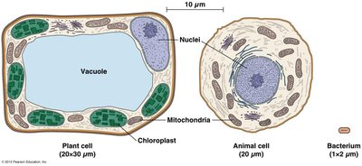
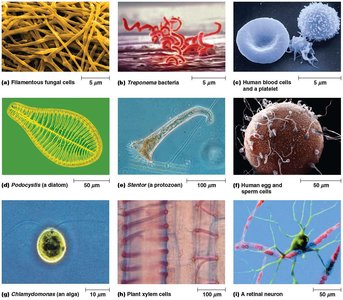
Understanding cell structure requires familiarity with units of measurement used in cell biology:
Micrometer (μm): 1 μm = 10-6 meters. Used for cells and organelles.
Nanometer (nm): 1 nm = 10-9 meters. Used for molecules and subcellular structures.
Angstrom (Å): 1 Å = 0.1 nm. Used for atomic-scale measurements, such as within proteins and DNA.
Microscopy: Tools for Studying Cells
Light Microscopy
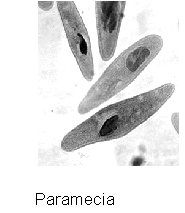
The light microscope was the first tool for cytologists, enabling the identification of major organelles. Brightfield microscopy, where white light passes through a specimen, is the most basic form. Samples are typically dead, fixed, and stained, which can introduce artifacts.
Microtome: Device for slicing thin sections (4–10 μm) of tissue for microscopy.
Staining: Dyes such as hematoxylin and eosin (H&E) or immunostaining are used to enhance contrast and identify specific molecules.
Optical Resolution
Resolution is the ability to distinguish two points as separate. It depends on the quality of the microscope's lenses. Higher resolution allows for greater detail.
Limit of Resolution: For light microscopes, typically 200–350 nm.
Magnification: Increases the apparent size of objects but does not improve detail beyond the resolution limit.
Types of Light Microscopes
Several types of light microscopes are used in cell biology:
Dissecting Microscope: Low magnification, used for larger specimens.
Compound Microscope: High magnification, used for thin sections of cells and tissues.
Inverted Compound Microscope: Used for observing living cultures in dishes.
Specialized Light Microscopy Techniques
To observe living cells and increase contrast, specialized techniques have been developed:
Phase-Contrast Microscopy: Enhances contrast by exploiting differences in refractive index.
Differential Interference Contrast (DIC) Microscopy: Uses polarized light for 3D-like images.
Fluorescence Microscopy: Detects fluorescent dyes or proteins to localize specific molecules.
Confocal Microscopy: Uses lasers to obtain sharp, optical sections and 3D reconstructions.
Fluorescence and Confocal Microscopy
Fluorescence microscopy uses fluorescent dyes or proteins (e.g., GFP) to visualize specific cellular components. Antibodies can be labeled with fluorescent tags to detect target molecules. Confocal microscopy allows for the collection of sharp, 2D images from different planes, which can be combined into 3D images.
Antibody: Protein that binds specifically to an antigen (target molecule).
GFP (Green Fluorescent Protein): Used to study protein localization in living cells.
Electron Microscopy
Principles and Advantages
Electron microscopes, developed in the 1930s, use beams of electrons instead of light, achieving much higher resolution (0.1–0.2 nm) and magnification (up to 100,000x). This has enabled visualization of subcellular structures and even individual atoms.
Transmission Electron Microscopy (TEM): Electrons pass through thin specimens to reveal internal structures.
Scanning Electron Microscopy (SEM): Electrons scan the surface, providing detailed 3D images of specimen surfaces.
Summary Table: Comparison of Light and Electron Microscopy
Feature | Light Microscopy | Electron Microscopy |
|---|---|---|
Source of Illumination | Visible light | Electron beam |
Resolution Limit | 200–350 nm | 0.1–0.2 nm |
Maximum Magnification | ~1,000–2,000x | Up to 100,000x |
Sample Preparation | Simple, can use living or fixed cells | Complex, samples must be fixed and dehydrated |
Visualization | Whole cells, organelles | Subcellular structures, molecules, atoms |
Key Equations
Micrometer to meter:
Nanometer to meter:
Angstrom to meter:
Conclusion
The development of cell theory and advances in microscopy have been fundamental to our understanding of cell biology. Modern techniques continue to reveal the complexity and beauty of the cellular world, forming the basis for all biological sciences.
