 Indietro
Chapter 3: Macromolecules – Proteins, Nucleic Acids, and Polysaccharides
Guida di studio - Note intelligenti
 Appunti personalizzati basati sui tuoi materiali, ampliati con definizioni chiave, esempi e contesto.
Macromolecules in Cell Biology
Introduction
Macromolecules are large, complex molecules essential for life. The four major classes—proteins, nucleic acids, polysaccharides, and lipids—play critical roles in cellular structure and function. This chapter focuses on the structure, function, and assembly of proteins, nucleic acids, and polysaccharides, which are foundational to cell biology.
Proteins
Classes and Functions of Proteins
Enzymes: Catalyze biochemical reactions, increasing reaction rates.
Structural proteins: Provide physical support and shape to cells and tissues.
Motility proteins: Involved in cellular movement and contraction.
Regulatory proteins: Control and coordinate cellular functions.
Transport proteins: Move substances across cellular membranes.
Signaling proteins: Facilitate communication between cells.
Receptor proteins: Allow cells to respond to environmental stimuli.
Defensive proteins: Protect against disease (e.g., antibodies).
Storage proteins: Serve as reservoirs of amino acids.
The Central Dogma of Molecular Biology
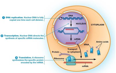
The central dogma describes the flow of genetic information: DNA is transcribed into RNA, which is then translated into protein. This process underlies all cellular activities.
Amino Acids: Structure and Properties
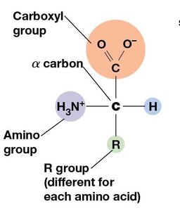
Proteins are polymers of amino acids. Each amino acid has a central (α) carbon, an amino group, a carboxyl group, a hydrogen atom, and a unique side chain (R group). The properties of amino acids are determined by their R groups.
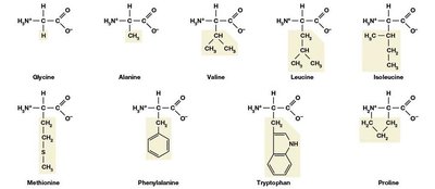
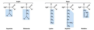
Non-polar (hydrophobic): Side chains are primarily hydrocarbons, making them water-insoluble.
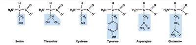
Polar, non-charged (hydrophilic): Side chains contain groups that form hydrogen bonds with water.
Polar, charged (hydrophilic): Side chains are either acidic (negatively charged) or basic (positively charged) at physiological pH.
Peptide Bond Formation and Directionality
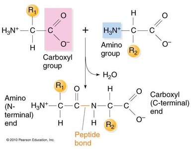
Amino acids are linked by peptide bonds formed via condensation (dehydration) reactions. The resulting polypeptide has directionality, with an N-terminus (amino end) and a C-terminus (carboxyl end). Sequences are always written from N- to C-terminus.
Protein Structure: Four Levels of Organization
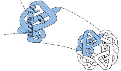
Primary structure: Linear sequence of amino acids in a polypeptide chain.
Secondary structure: Local folding patterns stabilized by hydrogen bonds, mainly α helices and β sheets.
Tertiary structure: Overall three-dimensional shape of a single polypeptide, stabilized by various interactions between R groups.
Quaternary structure: Association of multiple polypeptide subunits into a functional protein complex.
Primary Structure
The primary structure is the unique sequence of amino acids in a protein, which determines all higher levels of structure and function.

Secondary Structure: α Helix and β Sheet
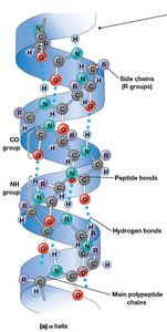
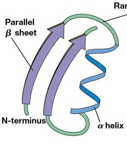
Secondary structures are stabilized by hydrogen bonds between backbone atoms. The α helix is a right-handed coil, while the β sheet consists of extended strands connected laterally by hydrogen bonds.
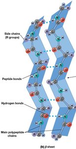
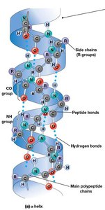
α Helix: 3.6 amino acids per turn; R groups project outward.
β Sheet: Strands can be parallel or antiparallel; R groups alternate above and below the plane.
Tertiary Structure: Interactions and Motifs
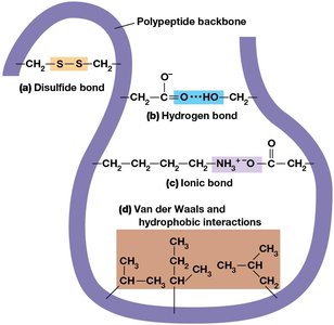
Tertiary structure is stabilized by:
Disulfide bonds: Covalent bonds between cysteine residues.
Hydrogen bonds: Between polar R groups.
Ionic bonds: Between charged R groups; sensitive to pH changes.
Van der Waals interactions: Weak attractions between nonpolar groups.
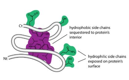
Hydrophobic interactions: Nonpolar side chains cluster away from water.
Protein Domains
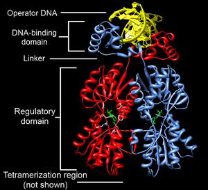
A domain is a discrete, locally folded unit of tertiary structure, often associated with a specific function (e.g., catalytic or DNA-binding domains). Proteins with multiple functions typically have multiple domains.
Quaternary Structure
Quaternary structure arises from the association of multiple polypeptide chains (subunits). The same types of bonds as in tertiary structure stabilize these assemblies. Example: Hemoglobin is a tetramer with two α and two β subunits.
Summary Table: Protein Structure Levels
Level of Structure | Basis of Structure | Kinds of Bonds and Interactions Involved |
|---|---|---|
Primary | Amino acid sequence | Covalent peptide bonds |
Secondary | Folding into α helix, β sheet, or random coil | Hydrogen bonds between NH and CO groups of peptide bonds in the backbone |
Tertiary | Three-dimensional folding of a single polypeptide chain | Disulfide bonds, hydrogen bonds, ionic bonds, van der Waals interactions, hydrophobic interactions |
Quaternary | Association of multiple polypeptides to form a multimeric protein | Same as for tertiary structure |
Fibrous, Globular, and Membrane Proteins
Fibrous proteins: Extended, filamentous structures (e.g., collagen, keratin).
Globular proteins: Compact, spherical shapes; most enzymes and regulatory proteins.
Membrane proteins: Associated with cellular membranes, often involved in transport or signaling.
Nucleic Acids
Structure and Function
Nucleic acids store, transmit, and express genetic information. DNA (deoxyribonucleic acid) and RNA (ribonucleic acid) are polymers of nucleotides.
Nucleotide Structure
Each nucleotide consists of a five-carbon sugar, a phosphate group, and a nitrogenous base (purine or pyrimidine).
Nucleic Acid Polymers
Nucleotides are linked by 3',5'-phosphodiester bonds via condensation reactions.
Polymers have directionality (5' to 3').
DNA forms a double helix with two antiparallel, complementary strands held together by hydrogen bonds between bases.
Polysaccharides
Structure and Function
Polysaccharides are long-chain polymers of monosaccharides, primarily serving structural and storage roles. The most common monosaccharide is glucose (C6H12O6).
Storage polysaccharides: Starch (plants), glycogen (animals, bacteria).
Structural polysaccharides: Cellulose (plant cell walls).
Key Concepts and Study Tips
Understand the relationship between structure and function for each macromolecule.
Be able to identify and describe the four levels of protein structure and the types of bonds involved.
Know the basic structure of amino acids, nucleotides, and monosaccharides.
Practice drawing and labeling diagrams of protein secondary structures and nucleic acid polymers.
