 Indietro
Enzymes: The Catalysts of Life – Comprehensive Study Notes
Guida di studio - Note intelligenti
 Appunti personalizzati basati sui tuoi materiali, ampliati con definizioni chiave, esempi e contesto.
Enzymes: The Catalysts of Life
Overview of Enzyme Catalysis
Enzymes are protein catalysts that facilitate nearly all cellular reactions. Their presence determines whether a reaction can occur under cellular conditions. The substrate is the molecule upon which an enzyme acts, binding to the enzyme's active site to undergo transformation.
Activation Energy and the Metastable State
Activation Energy Barrier
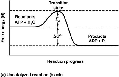
Many thermodynamically feasible reactions in cells do not proceed at appreciable rates due to the activation energy (EA) barrier. Activation energy is the minimum energy required for reactants to reach the transition state and form products. For example, ATP hydrolysis has a negative ΔGº', but ATP remains stable in water for days without catalysis.
Transition State: An intermediate stage with higher free energy than reactants.
Metastable State: Reactants are thermodynamically unstable but lack sufficient EA to react.
Overcoming the Activation Energy Barrier
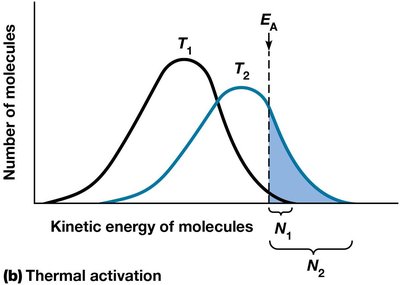
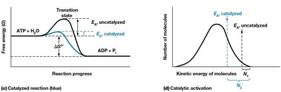
Reactions can be accelerated by increasing molecular energy (e.g., heat), but cells are isothermal. Catalysts, including enzymes, lower the EA requirement, allowing more molecules to react at cellular temperatures.
Enzymes as Biological Catalysts
Properties of Catalysts
Increase reaction rates by lowering EA.
Form transient, reversible complexes with substrates.
Change the rate at which equilibrium is achieved, not the equilibrium position.
Most enzymes are proteins, but some RNA molecules (ribozymes) also have catalytic activity.
The Active Site
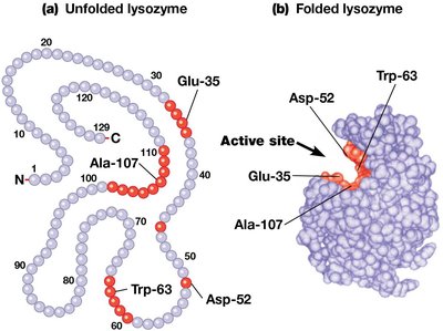
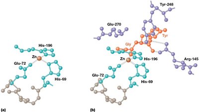
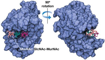
The active site is a cluster of amino acids forming a groove or pocket where substrates bind with high affinity. Only a few amino acids (cysteine, histidine, serine, aspartate, glutamate, lysine) are typically involved in active sites, participating in substrate binding and catalysis.
Prosthetic Groups
Prosthetic groups are nonprotein cofactors (often metal ions or coenzymes) tightly bound to the active site, essential for enzyme function. They frequently act as electron acceptors and explain nutritional requirements for vitamins and trace metals.
Enzyme Specificity
Enzymes exhibit high substrate specificity due to the shape and chemistry of their active sites. Some enzymes show group specificity, accepting substrates with common features, especially in polymer synthesis or degradation.
Enzyme Diversity and Nomenclature
Enzymes are named based on substrate or function and classified into six major classes:
Oxidoreductases: Electron transfer (e.g., alcohol dehydrogenase)
Transferases: Transfer functional groups (e.g., hexokinase)
Hydrolases: Hydrolytic cleavage (e.g., glucose-6-phosphatase)
Lyases: Addition/removal of groups (e.g., pyruvate decarboxylase)
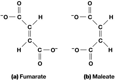
Isomerases: Functional group movement within a molecule (e.g., maleate isomerase)
Ligases: Joining two molecules (e.g., pyruvate carboxylase)
Sensitivity of Enzymes to Environmental Factors
Temperature Sensitivity
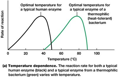
Enzyme activity increases with temperature due to increased kinetic energy, but excessive heat causes denaturation. Optimal temperature varies by organism; human enzymes peak at 37°C, while thermophilic bacteria enzymes function at higher temperatures.
pH Sensitivity
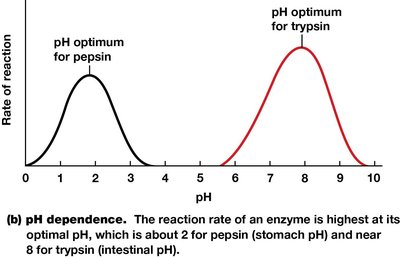
Most enzymes are active within a pH range of 3–4 units, with optimal pH depending on the enzyme and environment. Changes in pH affect charged amino acids, disrupting bonds and enzyme activity.
Sensitivity to Other Factors
Enzymes are also sensitive to inhibitors, activators, and ionic strength, which affect their tertiary structure and activity.
Substrate Binding, Activation, and Catalysis
Substrate Binding
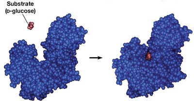
Substrates bind to the active site in the correct orientation, usually via hydrogen or ionic bonds. Binding is reversible and highly specific.
Induced-Fit Model
Unlike the rigid lock-and-key model, the induced-fit model describes how substrate binding induces a conformational change in both enzyme and substrate, optimizing the active site for catalysis.
Substrate Activation Mechanisms
Bond distortion: Makes bonds more susceptible to attack.
Proton transfer: Increases substrate reactivity.
Electron transfer: Forms temporary covalent bonds between enzyme and substrate.
The Catalytic Event
Substrate randomly collides and binds to the active site.
Binding induces conformational change, facilitating conversion to products.
Products are released; enzyme returns to original conformation.
Ribozymes: Catalytic RNA Molecules
Discovery and Examples
Ribozymes are RNA molecules with catalytic activity, discovered in the 1980s. Examples include self-splicing RNA in Tetrahymena thermophila and ribonuclease P, which processes tRNA precursors. Ribosomal RNA (rRNA) also acts as a catalyst in peptide bond formation.
Enzyme Kinetics
Quantitative Aspects of Catalysis
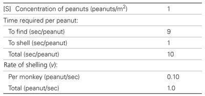
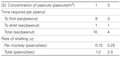
Enzyme kinetics studies the rate of substrate conversion to products, influenced by substrate, product, and inhibitor concentrations. Analogies, such as monkeys shelling peanuts, help illustrate kinetic principles.
[S] Concentration (peanuts/m2) | Time to find (sec/peanut) | Time to shell (sec/peanut) | Total (sec/peanut) | Rate per monkey (peanut/sec) | Total rate (peanut/sec) |
|---|---|---|---|---|---|
1 | 9 | 1 | 10 | 0.10 | 1.0 |
[S] Concentration (peanuts/m2) | Time to find (sec/peanut) | Time to shell (sec/peanut) | Total (sec/peanut) | Rate per monkey (peanut/sec) | Total rate (peanut/sec) |
|---|---|---|---|---|---|
1 | 9 | 1 | 10 | 0.10 | 1.0 |
3 | 3 | 1 | 4 | 0.25 | 2.5 |
Michaelis–Menten Kinetics
Most enzymes follow Michaelis–Menten kinetics, where initial velocity (v0) depends on substrate concentration ([S]). At low [S], v0 increases proportionally; at high [S], v0 approaches a maximum (Vmax), demonstrating saturation.
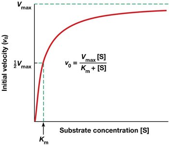
The Michaelis–Menten equation:
Km: Substrate concentration at half-maximal velocity.
Vmax: Maximum velocity at saturating substrate concentrations.
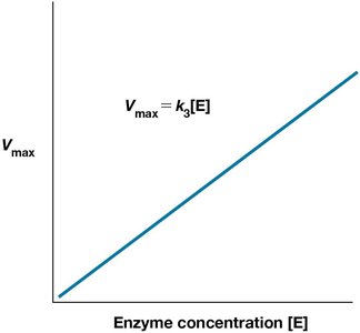
Vmax and Enzyme Concentration
Vmax is directly proportional to enzyme concentration. Increasing enzyme concentration increases Vmax.
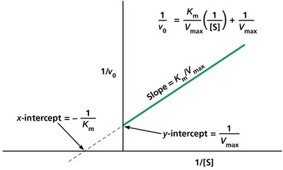
Lineweaver–Burk Double-Reciprocal Plot
The Lineweaver–Burk plot linearizes the Michaelis–Menten equation, allowing easier determination of Km and Vmax:
Enzyme Inhibition
Types of Inhibition
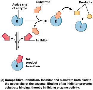
Enzyme inhibitors can act irreversibly (covalent binding, permanent loss of activity) or reversibly (noncovalent binding, dissociable). Reversible inhibitors are classified as competitive or noncompetitive.
Competitive Inhibitors: Bind the active site, competing with substrate. Vmax unchanged, Km increases.
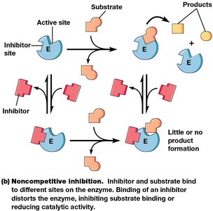
Noncompetitive Inhibitors: Bind elsewhere, causing conformational changes. Vmax decreases, Km may be unchanged or increased (mixed inhibition).
Enzyme Regulation
Substrate-Level Regulation
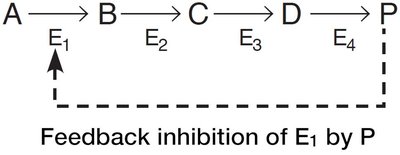
Enzyme rates are adjusted by substrate and product concentrations. Increased substrate raises reaction rates; increased product lowers them.
Allosteric Regulation and Covalent Modification
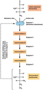
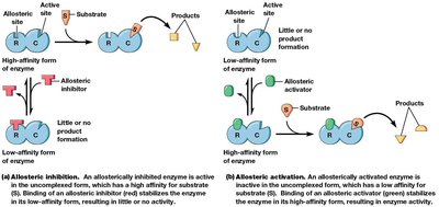
Allosteric regulation and covalent modification are key mechanisms for turning enzymes on/off. Allosteric enzymes have two conformations, regulated by effectors binding to a regulatory site distinct from the active site. Effectors may be activators or inhibitors.
Cooperativity
Allosteric enzymes often exhibit cooperativity, where substrate binding alters affinity at other sites. Positive cooperativity increases affinity; negative decreases it.
Covalent Modification
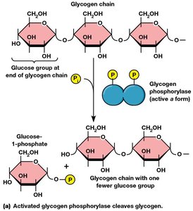
Enzyme activity can be regulated by addition/removal of chemical groups (phosphate, methyl, acetyl). Phosphorylation (by kinases) and dephosphorylation (by phosphatases) are common regulatory mechanisms.
Proteolytic Cleavage
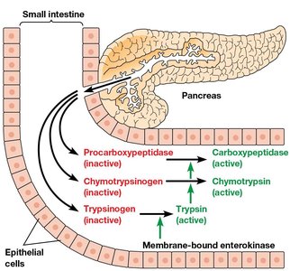
Some enzymes are activated by irreversible removal of part of the polypeptide chain (proteolytic cleavage). Pancreatic zymogens are synthesized in inactive form and activated by cleavage.
Summary Table: Major Classes of Enzymes
Class | Reaction Type | Example | Reaction Catalyzed |
|---|---|---|---|
Oxidoreductases | Oxidation-reduction | Alcohol dehydrogenase | Oxidation of ethanol to acetaldehyde |
Transferases | Transfer of functional groups | Hexokinase | Phosphorylation of glucose |
Hydrolases | Hydrolytic cleavage | Glucose-6-phosphatase | Cleavage of glucose-6-phosphate |
Lyases | Addition/removal of groups | Pyruvate decarboxylase | Removal of carboxyl group from pyruvate |
Isomerases | Isomerization | Maleate isomerase | Cis-trans isomerization of maleate |
Ligases | Joining molecules | Pyruvate carboxylase | Addition of CO2 to pyruvate |
Additional info: Academic context and explanations have been expanded for clarity and completeness. All images included are directly relevant to the adjacent content and reinforce key concepts.
