 Indietro
Macromolecules of the Cell: Structure and Function
Guida di studio - Note intelligenti
 Appunti personalizzati basati sui tuoi materiali, ampliati con definizioni chiave, esempi e contesto.
Macromolecules of the Cell
Introduction
Cells are composed of four major classes of macromolecules: proteins, nucleic acids, polysaccharides, and lipids. Each class plays a distinct and essential role in cellular structure and function.
Proteins
Functions of Proteins
Proteins are versatile macromolecules that perform a wide variety of functions in the cell:
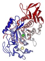
Enzymes: Catalyze biochemical reactions, increasing reaction rates without being consumed.
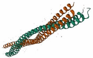
Structural proteins: Provide support and shape to cells and tissues (e.g., collagen, keratin).
Motility proteins: Involved in movement within cells and of the cell itself (e.g., actin, myosin).
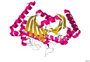
Regulatory proteins: Control cellular processes, including gene expression and cell cycle progression.
Transport proteins: Move molecules across membranes or within the cell (e.g., hemoglobin, ion channels).
Signaling proteins: Transmit signals to coordinate cellular activities (e.g., hormones, receptors).
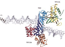
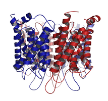
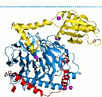
Receptor proteins: Receive and transmit signals from the environment or other cells.
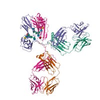
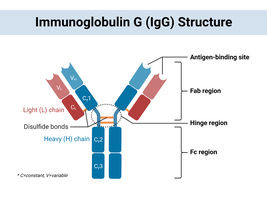
Defensive proteins: Protect the organism from disease (e.g., antibodies).
Storage proteins: Store amino acids or other substances for later use (e.g., ferritin).
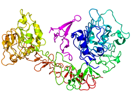
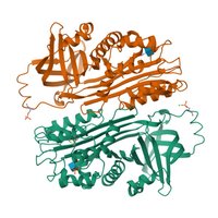
Amino Acids: The Building Blocks of Proteins
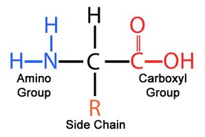
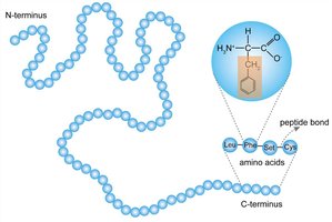
Proteins are polymers of amino acids. Each amino acid contains a central carbon (alpha carbon) bonded to an amino group, a carboxyl group, a hydrogen atom, and a variable side chain (R group).
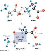
Amino acids are linked by peptide bonds to form polypeptides.
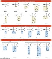
Classification of Amino Acids
Nonpolar (hydrophobic)
Polar uncharged
Positively charged (basic)
Negatively charged (acidic)
Protein Structure: Hierarchical Organization
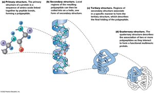
The function of a protein depends on its three-dimensional structure, which is organized into four levels:
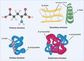
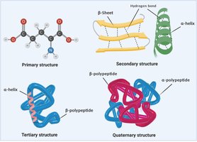
Primary structure: The linear sequence of amino acids in a polypeptide chain.
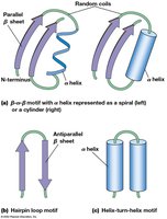
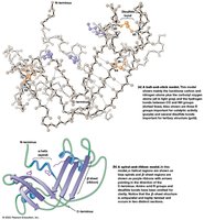
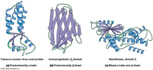
Secondary structure: Local folding patterns stabilized by hydrogen bonds (e.g., alpha helices, beta sheets).
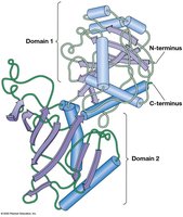
Tertiary structure: The overall three-dimensional shape formed by interactions among R groups.
Quaternary structure: The association of multiple polypeptide chains into a functional protein complex (not all proteins have this level).
Primary Structure
The primary structure is simply the sequence of amino acids in a protein, determined by the gene encoding the protein.
Secondary Structure
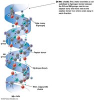
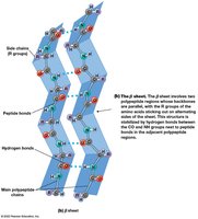
Secondary structure arises from hydrogen bonding between the backbone NH and CO groups. The two most common types are:
Alpha helix (α-helix): A right-handed coil stabilized by hydrogen bonds.
Beta sheet (β-sheet): Sheet-like arrangement formed by hydrogen bonds between parallel or antiparallel strands.
Tertiary Structure
Tertiary structure is the overall 3D shape of a single polypeptide, stabilized by interactions among R groups, including hydrogen bonds, ionic bonds, hydrophobic interactions, and van der Waals forces.
Quaternary Structure
Quaternary structure results from the association of two or more polypeptide chains (subunits) to form a functional protein complex.
Stabilizing Interactions in Protein Structure
Disulfide bonds: Covalent bonds between cysteine residues, important for stabilizing tertiary and quaternary structures.
Noncovalent interactions: Hydrogen bonds, ionic bonds, van der Waals interactions, and hydrophobic interactions.
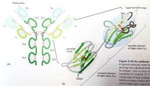
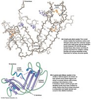
Fibrous and Globular Proteins
Proteins can be classified based on their shape and function:
Fibrous proteins: Elongated, structural proteins (e.g., collagen, keratin).
Globular proteins: Compact, functional proteins (e.g., enzymes, antibodies).
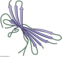
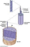
Case Study: Hemoglobin and Sickle Cell Disease
Hemoglobin is a quaternary protein responsible for oxygen transport in the blood. Sickle cell disease is caused by a single amino acid substitution (glutamic acid to valine) in the beta chain of hemoglobin, altering its interactions and causing aggregation.
Glutamic acid: Hydrophilic, forms ionic and hydrogen bonds.
Valine: Hydrophobic, promotes aggregation in deoxygenated hemoglobin.
*Additional info: Sickle cell hemoglobin forms fibers that distort red blood cells, leading to disease symptoms.*
Nucleic Acids
Structure and Function
Nucleic acids store, transmit, and express genetic information. The two main types are DNA (deoxyribonucleic acid) and RNA (ribonucleic acid). Their monomers are nucleotides, which consist of a sugar, a phosphate group, and a nitrogenous base (purine or pyrimidine).
ATP: The Energy Currency
ATP (adenosine triphosphate) is a nucleotide that stores and transfers energy in cells.
Nucleotide Polymerization
Nucleotides are linked by 3',5' phosphodiester bonds between the 3' hydroxyl of one sugar and the 5' phosphate of the next.
Base Pairing and DNA Structure
A pairs with T (or U in RNA)
G pairs with C
DNA forms a double helix structure, stabilized by hydrogen bonds between complementary bases.
Polysaccharides
Structure and Function
Polysaccharides are long chains of sugars or sugar derivatives. They serve as energy storage (e.g., starch, glycogen) or structural components (e.g., cellulose, chitin, peptidoglycan).
Monomer: Monosaccharides (simple sugars, e.g., glucose)
General formula: (e.g., for glucose)
Storage Polysaccharides
Starch: Main storage polysaccharide in plants (alpha-glycosidic bonds).
Glycogen: Main storage polysaccharide in animals (alpha-glycosidic bonds).
Structural Polysaccharides
Cellulose: Plant cell walls (beta-glycosidic bonds).
Chitin: Fungi and insect exoskeletons.
Peptidoglycan: Bacterial cell walls (contains amino acids).
Lipids
Structure and Function
Lipids are hydrophobic molecules, some of which are amphipathic (having both hydrophobic and hydrophilic regions). They function in energy storage, membrane structure, and signal transduction.
Fatty Acids
Fatty acids are long hydrocarbon chains with a carboxyl group. They can be:
Saturated: No double bonds (solid at room temperature).
Unsaturated: One or more double bonds (liquid at room temperature).
Triacylglycerols
Triacylglycerols (triglycerides) are storage lipids composed of glycerol linked to three fatty acids.
Phospholipids
Phospholipids are major components of cell membranes, consisting of a glycerol backbone, two fatty acids, and a phosphate group.
Steroids
Steroids are lipids with a characteristic four-ring structure, including cholesterol and steroid hormones.
