 Indietro
Membrane Transport and Osmosis: Mechanisms and Applications in Cell Biology
Guida di studio - Note intelligenti
 Appunti personalizzati basati sui tuoi materiali, ampliati con definizioni chiave, esempi e contesto.
Membrane Transport: Principles and Applications
Surface Area to Volume Ratio in Cells
The surface area to volume (SA/V) ratio is a fundamental concept in cell biology, influencing a cell's ability to exchange materials with its environment. Smaller cells or those with higher SA/V ratios are generally more efficient at nutrient uptake and waste removal.
Calculation: For a cubic cell, surface area = 6 × (side length)2; volume = (side length)3. For a rectangular cell, surface area = 2(lw + lh + wh); volume = l × w × h.
Implications: Cells with lower SA/V ratios face challenges in exchanging substances efficiently.
Adaptations: Larger cells may develop specialized structures or mechanisms to cope with reduced exchange capacity.
Example: Organism 1 (cubic, 1.5 µm sides) has a higher SA/V ratio than Organism 2 (rectangular, 2 × 5 × 2 µm), making Organism 2 less efficient in environmental interaction.
Types of Membrane Transport
Passive Transport
Passive transport involves the movement of molecules along their concentration or electrochemical gradient, requiring no energy input from the cell.
Simple Diffusion: Direct movement of small, non-polar molecules across the lipid bilayer.
Facilitated Diffusion: Movement of molecules via transport proteins, allowing larger or polar substances to cross membranes.
Example: Oxygen and carbon dioxide diffuse across cell membranes by simple diffusion; glucose and ions often require facilitated diffusion.
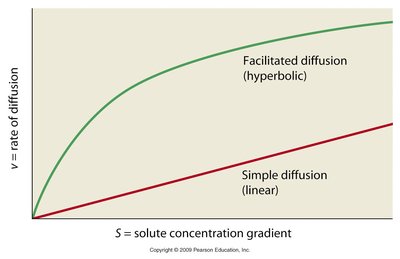
Additional info: Facilitated diffusion shows a hyperbolic relationship with solute concentration, while simple diffusion is linear.
Osmosis and Tonicity
Osmosis is the diffusion of water across a selectively permeable membrane. Tonicity describes the relative concentration of solutes outside versus inside the cell, affecting water movement.
Hypotonic: Lower solute concentration outside; water enters the cell.
Hypertonic: Higher solute concentration outside; water leaves the cell.
Isotonic: Equal solute concentration; no net water movement.
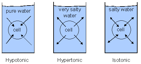
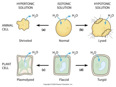
Example: Animal cells in hypotonic solutions may lyse, while plant cells become turgid. In hypertonic solutions, animal cells shrivel and plant cells undergo plasmolysis.
Microscopic Observations of Osmosis
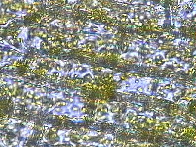
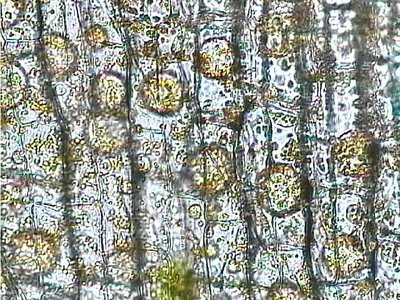
Microscopy can reveal the effects of osmotic conditions on plant cells, such as Elodea cells.
Isotonic: Cells retain normal shape.
Hypertonic: Cells undergo plasmolysis, with cytoplasm shrinking away from the cell wall.
Facilitated Transport and Transport Proteins
Transport Systems in the Plasma Membrane
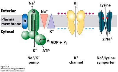
Cells utilize various transport proteins to move substances across membranes, including channels, carriers, and pumps.
Channels: Allow passive movement of ions or water.
Carriers: Bind and transport specific molecules.
Pumps: Use energy to move substances against gradients.
Types of Transporters: Uniport, Symport, Antiport
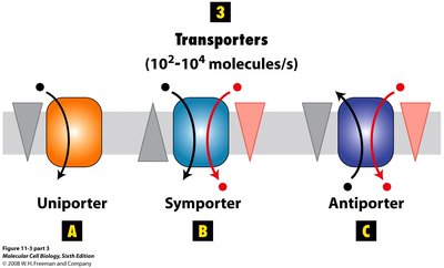
Transport proteins can be classified based on the direction and number of substances they move.
Uniport: Transports one substance in one direction.
Symport: Transports two substances in the same direction.
Antiport: Transports two substances in opposite directions.
Ion Channels and Gated Transport
Ion Channels and Their Regulation
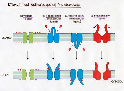
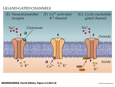
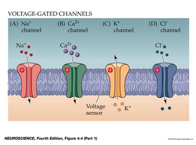
Ion channels are integral membrane proteins that allow ions to pass through the membrane. Their activity is regulated by various stimuli.
Voltage-gated: Open in response to changes in membrane potential.
Ligand-gated: Open when a specific molecule binds.
Mechanically-gated: Open in response to physical deformation.
Electrophysiology Techniques
Patch Clamp and Voltage Clamp Methods
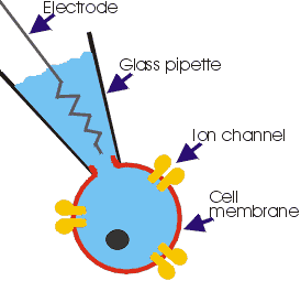
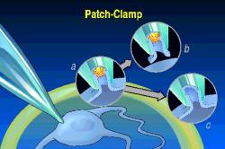
Electrophysiology techniques are used to study ion channel function and membrane transport.
Patch Clamp: Measures currents through individual ion channels.
Voltage Clamp: Maintains membrane potential to study ion flow.
Active Transport and ATPases
Energy Sources for Membrane Transport
Active transport requires energy, often supplied by ATP hydrolysis or ion gradients.
ATPases: Enzymes that hydrolyze ATP to drive transport.
Indirect Active Transport: Uses the gradient of one ion to transport another against its gradient.
Types of ATPases
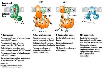
ATPases are classified based on their structure, location, and function.
Type | Location | Function |
|---|---|---|
P-class | Plasma membrane of plants, fungi, bacteria, higher eukaryotes | Na+/K+ pump, Ca2+ pump. H+ pump. ions |
V-class | Vacuolar membranes, endo- somal/lysosomal membranes | Proton transport. and |
F-class | Bacterial plasma membrane, mitochondria, chloroplasts | ATP- synthesis. to make atp? can they work as atpases? |
ABC superfamily | Various membranes | Transport of diverse abc for small molecules using atp , one exception |
Example: The Na+/K+ ATPase maintains cellular ion gradients essential for cell function.
Additional info: ABC transporters are involved in multidrug resistance and cystic fibrosis.
Sodium binding: Three intracellular Na⁺ ions attach to the pump protein from inside the cell.
ATP hydrolysis: ATP breaks down into ADP, transferring a phosphate group to the pump (phosphorylation).
Sodium release: The pump changes shape, opening to the outside and releasing the three Na⁺ ions into the extracellular fluid.
Potassium binding: Two extracellular K⁺ ions attach to the newly exposed binding sites on the outside of the pump.
Dephosphorylation: The phosphate group splits off the pump, causing it to shift back to its original shape.
Potassium release: The pump opens to the inside of the cell and releases the two K⁺ ions into the cytoplasm, readying the pump for a new cycle. 3 sodium's out 2 potassium in . negative charge inside.
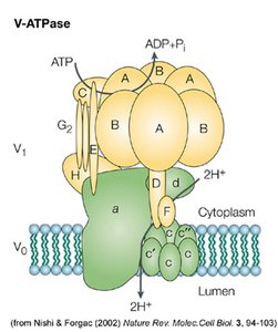
V TYPE ATPase
