 Indietro
Polysaccharides: Structure, Function, and Classification in Cell Biology
Guida di studio - Note intelligenti
 Appunti personalizzati basati sui tuoi materiali, ampliati con definizioni chiave, esempi e contesto.
The Macromolecules of the Cell
Polysaccharides: Structure and Function
Polysaccharides are essential macromolecules in cell biology, serving both structural and energy storage roles. They are polymers composed of repeating sugar units, typically monosaccharides, and their structure and function are determined by the types of sugars and the bonds linking them.
Definition: Polysaccharides are long-chain polymers of sugars and sugar derivatives.
Repeating Units: Usually consist of a single kind of repeating unit (e.g., glucose in starch), or an alternating pattern of two kinds (e.g., glucose and fructose in sucrose).
Roles: Serve in cellular structures (e.g., cellulose in plants) and energy storage (e.g., starch, glycogen).
Oligosaccharides: Short polymers sometimes attached to cell surface proteins, playing a role in cellular recognition.
Monosaccharides: The Building Blocks
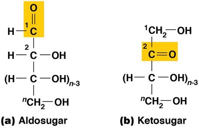
Monosaccharides are the monomeric units of polysaccharides. They are classified based on their carbonyl group and the number of carbon atoms.
Aldosugars: Sugars with a terminal carbonyl group (aldehyde).
Ketosugars: Sugars with an internal carbonyl group (ketone).
Classification by Carbon Number:
Trioses (3 carbons)
Tetroses (4 carbons)
Pentoses (5 carbons) – e.g., ribose in RNA, deoxyribose in DNA
Hexoses (6 carbons) – e.g., glucose
Heptoses (7 carbons)
Glucose: The Most Common Monosaccharide
D-glucose (C6H12O6) is the most prevalent monosaccharide in cells. Its formula, CnH2nOn, led to the term "carbohydrate" as "hydrates of carbon." The carbons are numbered from the more oxidized, carbonyl end.
Photosynthesis: For every CO2 incorporated into a sugar, one H2O is added.
Numbering: Carbons are numbered from the carbonyl end.
Stereoisomers of Glucose
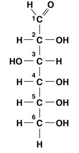
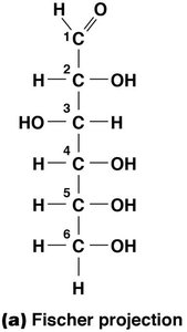
Glucose has four asymmetric carbon atoms, resulting in 16 possible stereoisomers. D-glucose is the most stable and biologically relevant form.
Asymmetric Carbons: Carbons 2, 3, 4, and 5.
Number of Stereoisomers:
D-glucose: Most stable and common in nature.
Ring Structure of D-Glucose
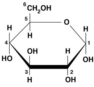
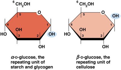
D-glucose exists in equilibrium between linear and ring forms. The ring form, called a pyranose ring, is more stable and is depicted in the Haworth projection. The ring is formed when the oxygen atom of the hydroxyl group on carbon 5 bonds with carbon 1.
Pyranose Ring: Five carbon atoms and one oxygen atom.
Equilibrium: Linear and ring forms coexist in cells.
Alpha and Beta Ring Forms
The ring formation of D-glucose can result in two forms, α and β, depending on the spatial orientation of the hydroxyl group on carbon 1.
α Form: Hydroxyl group downward.
β Form: Hydroxyl group upward.
Disaccharides: Covalent Linkage of Monosaccharides
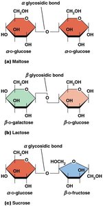
Disaccharides are formed by the covalent linkage of two monosaccharides via a glycosidic bond, which is created by a condensation reaction (elimination of water).
Common Disaccharides:
Maltose: Two glucose units (α glycosidic bond)
Lactose: One glucose and one galactose (β glycosidic bond)
Sucrose: One glucose and one fructose (α glycosidic bond)
Bond Types: α glycosidic (e.g., maltose), β glycosidic (e.g., lactose)
Storage Polysaccharides: Starch and Glycogen
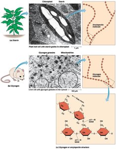
Starch and glycogen are the primary storage polysaccharides in plants and animals, respectively. Both are composed of α-D-glucose units linked by α(1→4) glycosidic bonds, with occasional α(1→6) bonds for branching.
Starch: Found in plant tissue; consists of unbranched amylose (10–30%) and branched amylopectin (70–90%). Amylopectin has α(1→6) branches every 12–25 glucose units.
Glycogen: Highly branched, with branches every 8–10 glucose units; stored in liver and muscle tissues in animals, and in bacteria.
Enzymatic Breakdown: Starch is broken down by amylase and maltase.
Structural Polysaccharides: Cellulose
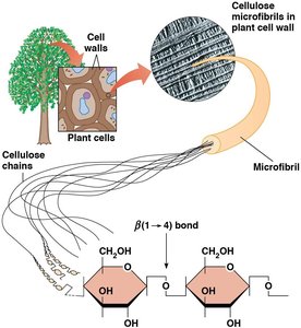
Cellulose is the most abundant structural polysaccharide in plants, composed of repeating β-D-glucose monomers. Its β(1→4) linkages result in rigid, linear rods that aggregate into microfibrils, providing structural support to plant cell walls.
Cellulose: Found in plant cell walls; mammals cannot digest it.
Microfibrils: Aggregates of cellulose chains, 5–20 nm in diameter.
Matrix: Plant and fungal cell walls contain microfibrils in a matrix of hemicellulose, pectin, and extensin protein.
Other Structural Polysaccharides: Fungi, Bacteria, and Insects
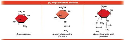
Structural polysaccharides vary among organisms. Fungal cell walls may contain β(1→4) or β(1→3) linkages. Bacterial cell walls are composed of alternating GlcNAc (N-acetylglucosamine) and MurNAc (N-acetylmuramic acid) units. Chitin, found in insect exoskeletons and fungal cell walls, consists of GlcNAc units joined by β(1→4) bonds.
Bacterial Cell Walls: Alternating GlcNAc and MurNAc units.
Chitin: GlcNAc units only, joined by β(1→4) bonds; found in insects, crustaceans, and fungi.
Polysaccharide Structure and Glycosidic Bonds
The type of glycosidic bond (α or β) determines the structure and properties of polysaccharides.
α Polysaccharides: Starch and glycogen form loose helices, not highly ordered due to side chains.
β Polysaccharides: Cellulose forms rigid, linear rods that aggregate into microfibrils.
Cell Wall Matrix: Plant and fungal cell walls contain microfibrils in a matrix of other polymers and proteins.
Comparison Table: Storage vs. Structural Polysaccharides
Type | Monomer | Bond Type | Structure | Function |
|---|---|---|---|---|
Starch | α-D-glucose | α(1→4), α(1→6) | Helical, branched | Energy storage (plants) |
Glycogen | α-D-glucose | α(1→4), α(1→6) | Highly branched | Energy storage (animals, bacteria) |
Cellulose | β-D-glucose | β(1→4) | Linear, microfibrils | Structural (plants) |
Chitin | GlcNAc | β(1→4) | Linear | Structural (insects, fungi) |
Bacterial Cell Wall | GlcNAc, MurNAc | β(1→4) | Alternating units | Structural (bacteria) |
Concept Check: Polysaccharides vs. Proteins and Nucleic Acids
Polysaccharides, proteins, and nucleic acids are all important macromolecules in cell structure and function. They are similar in being polymers of repeating units, but differ in their monomers, bond types, and biological roles.
Similarity: All are polymers with specific repeating units.
Difference: Polysaccharides are made of sugars, proteins of amino acids, nucleic acids of nucleotides.
Bond Types: Glycosidic (polysaccharides), peptide (proteins), phosphodiester (nucleic acids).
Function: Polysaccharides mainly for structure and energy storage; proteins for catalysis, structure, signaling; nucleic acids for information storage and transfer.
