 Indietro
Structure and Function of Biological Membranes
Guida di studio - Note intelligenti
 Appunti personalizzati basati sui tuoi materiali, ampliati con definizioni chiave, esempi e contesto.
Membrane Structure: Historical Discoveries
Development of Membrane Models
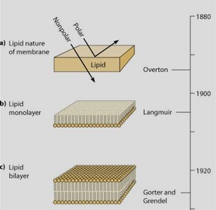
The understanding of biological membrane structure has evolved through key experiments and models. Early hypotheses proposed a lipid nature for membranes, which was later refined into the lipid bilayer model and the fluid mosaic model.
Overton (1895): Proposed that cell membranes are composed of lipid-like substances, allowing certain molecules to permeate.
Langmuir (1917): Demonstrated that lipids form monolayers on water, with hydrophilic heads facing water and hydrophobic tails away.
Gorter and Grendel (1925): Extracted lipids from red blood cells and found that the surface area of the extracted lipids was twice that of the cells, leading to the conclusion that membranes are lipid bilayers.
Davson and Danielli (1935): Proposed the "sandwich" model, with proteins coating both sides of the lipid bilayer.
Robertson (1960): Electron microscopy revealed a trilaminar (three-layered) appearance, supporting the unit membrane concept.
Singer and Nicolson (1972): Introduced the fluid mosaic model, describing membranes as a mosaic of proteins floating in a fluid lipid bilayer.

Membrane Composition
Protein, Lipid, and Carbohydrate Content
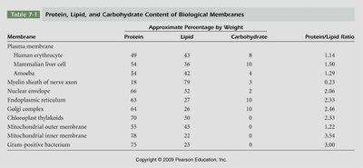
Biological membranes are composed of varying proportions of proteins, lipids, and carbohydrates, depending on the organism and membrane type. This composition determines membrane properties and functions.
Proteins: Involved in transport, signaling, and structural support.
Lipids: Form the bilayer, providing a barrier and matrix for proteins.
Carbohydrates: Usually attached to proteins or lipids, important for cell recognition and signaling.
Membrane | Protein (%) | Lipid (%) | Carbohydrate (%) | Protein/Lipid Ratio |
|---|---|---|---|---|
Plasma membrane | 49 | 43 | 8 | 1.14 |
Human erythrocyte | 54 | 42 | 8 | 1.29 |
Mammalian liver cell | 54 | 36 | 10 | 1.50 |
Amoeba | 18 | 77 | 5 | 0.23 |
Myelin sheath of nerve axon | 18 | 79 | 3 | 0.23 |
Nuclear envelope | 58 | 36 | 6 | 1.61 |
Endoplasmic reticulum | 64 | 32 | 4 | 2.00 |
Golgi complex | 64 | 32 | 4 | 2.00 |
Chloroplast thylakoids | 70 | 28 | 2 | 2.50 |
Mitochondrial outer membrane | 52 | 48 | 0 | 1.08 |
Mitochondrial inner membrane | 78 | 22 | 0 | 3.55 |
Gram positive bacterium | 75 | 25 | 0 | 3.00 |
Membrane Lipids: Structure and Dynamics
Lipid Bilayer and Fluidity
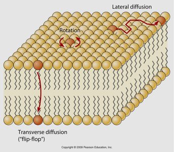
The lipid bilayer is the fundamental structure of biological membranes, composed mainly of phospholipids, cholesterol, and glycolipids. The bilayer is dynamic, allowing lateral and rotational movement of lipids, and occasionally, transverse diffusion (flip-flop) facilitated by enzymes such as flippases.
Lateral diffusion: Lipids move side-to-side within the same leaflet.
Rotation: Lipids rotate around their axis.
Transverse diffusion (flip-flop): Lipids move from one leaflet to the other, a process that is rare without enzymatic assistance.
Saturated vs. Unsaturated Fatty Acids
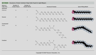
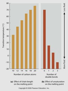
Fatty acids in membrane lipids can be saturated (no double bonds), unsaturated (one or more double bonds), or polyunsaturated (multiple double bonds). The degree of saturation affects membrane fluidity and phase transition temperature.
Saturated fatty acids: Pack tightly, making membranes less fluid and increasing transition temperature.
Unsaturated fatty acids: Introduce kinks, preventing tight packing, increasing fluidity, and lowering transition temperature.
Phase Transition and Differential Scanning Calorimetry
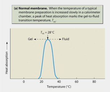
Membrane fluidity changes with temperature. The transition temperature (Tm) is the point at which the membrane shifts from a gel (ordered) to a fluid (disordered) state. Differential scanning calorimetry is used to measure this transition.
Higher Tm: More saturated fatty acids, longer chains.
Lower Tm: More unsaturated fatty acids, shorter chains.
Cholesterol and Membrane Microdomains
Role of Cholesterol
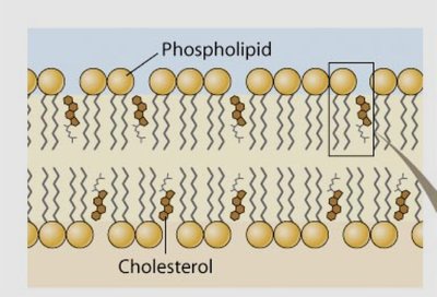
Cholesterol is an essential component of animal cell membranes, modulating membrane fluidity and stability. It interacts with phospholipids via hydrogen bonds and hydrophobic interactions.
At high temperatures: Cholesterol decreases membrane fluidity by restricting phospholipid movement.
At low temperatures: Cholesterol prevents membranes from becoming too rigid (gelling) by disrupting regular packing of phospholipids.
Lipid Rafts
Lipid rafts are specialized microdomains within the membrane, enriched in cholesterol, saturated fatty acids, and specific proteins. They serve as organizing centers for signaling molecules and influence membrane fluidity and protein trafficking.
Composition: High in cholesterol and sphingolipids.
Function: Facilitate cell signaling, protein sorting, and membrane trafficking.

Techniques to Study Membrane Structure
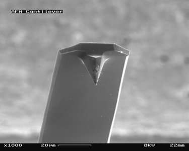
Advanced microscopy techniques, such as atomic force microscopy (AFM), allow visualization of membrane microdomains like lipid rafts. AFM uses a probe to scan the membrane surface, detecting physical and chemical interactions.
AFM probe: Interacts with the specimen via van der Waals forces, hydrogen bonds, and other interactions.
Applications: Imaging membrane topography, identifying lipid rafts, and studying protein-lipid interactions.
Membrane Proteins: Structure and Analysis
Types of Membrane Proteins
Membrane proteins are classified based on their association with the lipid bilayer:
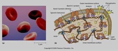
Integral (transmembrane) proteins: Span the bilayer, often with hydrophobic domains within the membrane and hydrophilic domains exposed to aqueous environments.
Peripheral proteins: Loosely attached to the membrane surface, often via interactions with integral proteins or lipid head groups.
Methods to Study Membrane Proteins
Several biochemical techniques are used to isolate and analyze membrane proteins:
Detergent solubilization: Detergents like SDS disrupt membranes, allowing extraction of proteins.
Gel electrophoresis: Proteins are separated based on size and charge after solubilization.
Staining: Coomassie blue binds to aromatic amino acids, arginine, and histidine, allowing visualization of proteins in gels.
Freeze-fracture electron microscopy: Reveals the distribution of proteins within the membrane by splitting the bilayer and imaging the exposed surfaces.

Functional Specialization of Membranes
Membrane Functions
Biological membranes perform several essential functions:
Barrier function: The lipid bilayer acts as a selective barrier, controlling the entry and exit of substances.
Transport and signaling: Membrane proteins mediate transport of ions and molecules, and participate in signal transduction.
Cell communication: Membranes contain proteins involved in cell-cell recognition, adhesion, and signaling.
Example Problem: Transmembrane Protein Topology
Problem Analysis
A transmembrane protein with 1000 amino acids has specific residues located in different regions:
Amino acid 5: External, interacts with aqueous environment (likely hydrophilic).
Amino acid 90: Within the membrane bilayer (likely hydrophobic).
Amino acids 100-600: Intracellular, with 200-400 forming a compact structure (likely hydrophilic, with some buried hydrophobic residues).
Amino acid 979: External, forms a weak ionic bond with Cl- (likely basic or positively charged).
Properties of these amino acids:
Hydrophilic (polar or charged): Found on surfaces exposed to aqueous environments (e.g., amino acids 5 and 979).
Hydrophobic (nonpolar): Found within the membrane bilayer (e.g., amino acid 90).
Charged (basic or acidic): Involved in ionic interactions (e.g., amino acid 979).
Additional info: The arrangement of amino acids in transmembrane proteins reflects the amphipathic nature of the membrane, with hydrophobic regions spanning the bilayer and hydrophilic regions exposed to the cytoplasm or extracellular space.
