 Indietro
Cell Division and Chromosome Heredity: Mitosis, Meiosis, and Sex Chromosome Inheritance
Guida di studio - Note intelligenti
 Appunti personalizzati basati sui tuoi materiali, ampliati con definizioni chiave, esempi e contesto.
Cell Division and Chromosome Heredity
Mitosis: Mechanism and Significance
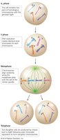
Mitosis is the process by which somatic cells divide to produce genetically identical daughter cells. It is essential for growth, development, and tissue repair in multicellular organisms.
Key Stages: Includes G1 phase (cell growth), S phase (DNA replication), G2 phase (preparation for division), metaphase (chromosomes align), and telophase (cell divides).
Genetic Identity: Daughter cells are genetically identical to the parent cell.
Example: Skin cell regeneration after injury.
Cell Cycle Checkpoints and Regulation
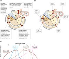
The cell cycle is tightly regulated by genetically controlled signals and checkpoints. These checkpoints ensure proper cell division and prevent errors that could lead to disease.
Checkpoints: G1, S, G2, and metaphase checkpoints monitor DNA integrity, chromosome attachment, and cell size.
Protein Interactions: Cyclins and cyclin-dependent kinases (CDKs) regulate progression through the cell cycle.
Cancer: Mutations in checkpoint genes can lead to uncontrolled cell proliferation, a hallmark of cancer.
Comparison of Mitosis and Meiosis
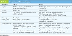
Mitosis and meiosis are two distinct types of cell division. Mitosis produces identical cells, while meiosis generates gametes for sexual reproduction, introducing genetic diversity.
Mitosis: One division, produces two identical diploid cells.
Meiosis: Two divisions, produces four genetically distinct haploid cells.
Homologous Chromosomes: Pairing and recombination occur only in meiosis.
Characteristic | Mitosis | Meiosis |
|---|---|---|
Purpose | Growth and repair | Sexual reproduction |
Location | Somatic cells | Germ-line cells |
Mechanics | One division | Two divisions |
Homologous chromosomes | Do not pair | Pair and recombine |
Sister chromatids | Separate in anaphase | Separate in anaphase II |
Product | Two diploid cells | Four haploid cells |
Meiosis: Mechanism and Genetic Consequences
Overview of Meiosis
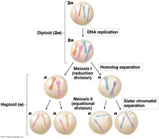
Meiosis is a specialized cell division process that produces gametes (sperm and eggs) for sexual reproduction. It involves one round of DNA replication followed by two cell divisions, resulting in four haploid cells.
Meiosis I: Homologous chromosomes separate, reducing chromosome number by half.
Meiosis II: Sister chromatids separate, similar to mitosis.
Genetic Diversity: Crossing over and independent assortment increase genetic variation.
Stages of Meiosis I
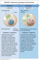
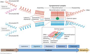
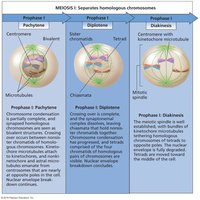
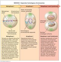
Meiosis I is divided into prophase I, metaphase I, anaphase I, and telophase I. Prophase I is further subdivided into leptotene, zygotene, pachytene, diplotene, and diakinesis, during which homologous chromosomes pair and recombine.
Prophase I: Homologs pair and recombine, forming the synaptonemal complex.
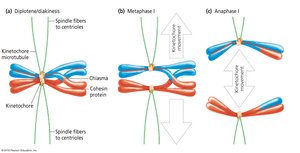
Metaphase I: Homologous pairs align at the metaphase plate.
Anaphase I: Homologs separate to opposite poles.
Telophase I: Cells divide, each with half the original chromosome number.
Sex Chromosomes and Pseudoautosomal Regions
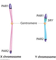
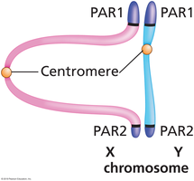
During meiosis, X and Y chromosomes pair via pseudoautosomal regions (PARs), allowing for proper segregation. These regions are present at both ends of the X and Y chromosomes.
Pseudoautosomal Regions: PAR1 and PAR2 facilitate pairing and recombination between X and Y.
SRY Gene: Located on the Y chromosome, critical for male sex determination.
Meiosis II: Separation of Sister Chromatids
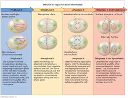
Meiosis II resembles mitosis, where sister chromatids are separated, resulting in four haploid cells. Each cell contains one chromosome from each homologous pair.
Prophase II: Chromosomes condense, spindle forms.
Metaphase II: Chromosomes align at the metaphase plate.
Anaphase II: Sister chromatids separate.
Telophase II: Cells divide, forming four haploid gametes.
Meiosis and Mendelian Ratios
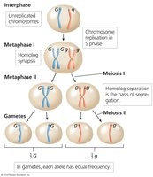
The separation of homologs and sister chromatids during meiosis provides the physical basis for Mendel's laws of segregation and independent assortment.
Law of Segregation: Each gamete receives one allele from each gene pair.
Law of Independent Assortment: Alleles of different genes assort independently during gamete formation.
Example: In a heterozygote (Aa), meiosis produces gametes with equal frequency of A and a alleles.
Chromosome Theory of Heredity and Sex Chromosome Inheritance
Chromosome Theory of Heredity
The chromosome theory of heredity, proposed by Sutton and Boveri, states that genes are carried on chromosomes and their behavior during meiosis explains inheritance patterns.
Experimental Evidence: Morgan's studies in Drosophila melanogaster validated the theory.
Wild Type: The most common phenotype in a population.
X-Linked Inheritance in Drosophila
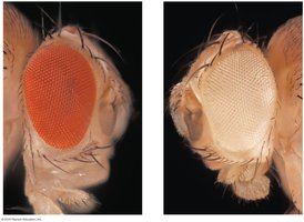
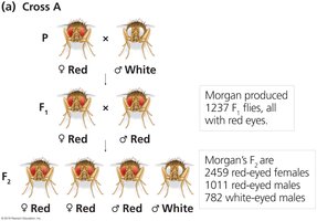
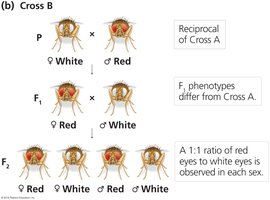
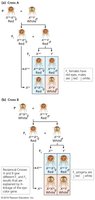
X-linked inheritance refers to genes located on the X chromosome. Morgan's experiments with white-eyed mutants in fruit flies demonstrated X-linked recessive inheritance.
White-Eyed Mutant: The allele for white eyes is recessive and located on the X chromosome.
Reciprocal Crosses: Showed different phenotypic ratios depending on the sex of the parent carrying the mutant allele.
Hemizygosity: Males have only one X chromosome, so any allele present is expressed.
Nondisjunction and Exceptional Progeny
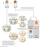
Nondisjunction is the failure of chromosomes to separate properly during meiosis, leading to abnormal chromosome numbers and unexpected phenotypes.
Exceptional Progeny: Females with two X chromosomes and one Y, or males with only one X and no Y, result from nondisjunction.
Example: Calvin Bridges observed rare white-eyed females and red-eyed males in crosses due to nondisjunction.
Sex Determination Mechanisms
Sex determination varies among species. In Drosophila, the X/autosome ratio determines sex, while in mammals, the presence of the SRY gene on the Y chromosome is critical.
X/A Ratio: Males have a ratio of 0.5, females have 1.0.
SRY Gene: Initiates male development in mammals.
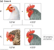
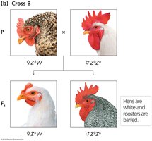
Other Systems: Birds and some reptiles use the Z/W system, where females are ZW and males are ZZ.
Human Sex-Linked Transmission and Dosage Compensation
Patterns of Sex-Linked Inheritance
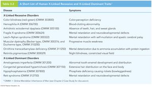
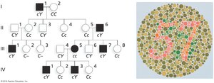
Human sex-linked traits follow distinct inheritance patterns. X-linked recessive traits are more frequently expressed in males, while X-linked dominant traits are expressed in both sexes. Y-linked traits are transmitted only from father to son.
X-Linked Recessive: Trait appears more often in males.
X-Linked Dominant: Trait appears in both sexes, but males are hemizygous.
Y-Linked: Exclusively male-to-male transmission.
Dosage Compensation Mechanisms
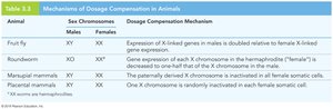
Dosage compensation equalizes the expression of sex-linked genes between males and females. Different organisms use distinct mechanisms to achieve this balance.
Mechanisms: Include doubling expression in males, downregulation in hermaphrodites, and X-chromosome inactivation in females.
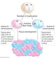
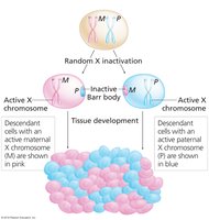
Lyon Hypothesis: Random X inactivation in female mammals leads to mosaicism.
Barr Body: The inactivated X chromosome forms a condensed structure visible in the nucleus.
Animal | Sex Chromosomes | Dosage Compensation Mechanism |
|---|---|---|
Fruit fly | XY/XX | Expression of X-linked genes in males is doubled |
Roundworm | XO/XX | Gene expression of each X is decreased in hermaphrodites |
Marsupial mammal | XY/XX | Paternally derived X is inactivated in females |
Placental mammal | XY/XX | One X is randomly inactivated in each female cell |
Random X-Chromosome Inactivation and Mosaicism
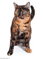
In placental mammals, random X inactivation results in females being mosaics, with some cells expressing the maternal X and others the paternal X. This phenomenon is visible in calico and tortoiseshell cats.
XIST Gene: Produces RNA that coats and inactivates the X chromosome.
Mosaicism: Leads to unique patterns of gene expression and visible traits.
Example: Calico cats display patches of orange and black fur due to X inactivation.
Mechanism of X Inactivation
X inactivation is not absolute; some genes escape inactivation. The XIST gene acts in cis, affecting only the chromosome from which it is transcribed.
Escape from Inactivation: 15-50% of genes may remain active on the inactivated X.
XIST RNA: Spreads across the chromosome, silencing gene expression.
Additional info: X inactivation is crucial for preventing dosage imbalance of X-linked genes, which could otherwise lead to developmental abnormalities.
