 Indietro
Cell Division: Mitosis and Meiosis – Genetics Study Guide
Guida di studio - Note intelligenti
 Appunti personalizzati basati sui tuoi materiali, ampliati con definizioni chiave, esempi e contesto.
Recap Lecture 1: Fundamental Concepts in Genetics
Gene and Allele
The gene is the basic unit of inheritance, encoding information for a specific trait. Alleles are alternative forms of a gene, which can result in different phenotypes.
Gene: Segment of DNA coding for a protein or functional RNA.
Allele: Variant form of a gene, e.g., A and a for a gene controlling flower color.
Example: The gene for eye color may have alleles for blue or brown eyes.
Genotype and Phenotype
The genotype refers to the complete set of genetic material or the specific alleles present in an organism. The phenotype is the observable traits resulting from the genotype.
Genotype: The genetic constitution (e.g., AA, Aa, aa).
Phenotype: Observable characteristics (e.g., tall or short plants).
Central Dogma of Molecular Biology
The central dogma describes the flow of genetic information: DNA is transcribed to mRNA, which is translated into protein.
DNA → mRNA → Protein
Example: The gene for hemoglobin is transcribed and translated to produce the hemoglobin protein.
Chromosome
A chromosome is a package of DNA containing part or all of the genetic material of an organism.
Chromatin: DNA-protein complex forming chromosomes.
Centromere: Region where sister chromatids are joined.
Forward vs. Reverse Genetic Analyses
Forward genetics: Start with a phenotype, identify the gene responsible.
Reverse genetics: Start with a gene, determine its function by observing phenotypes after manipulation.
Model Organisms
Model organisms are non-human species with stable genetic backgrounds used for biological research.
Examples: Drosophila melanogaster (fruit fly), Mus musculus (mouse).
Chapter 2: Mitosis and Meiosis
Cell Cycle Overview
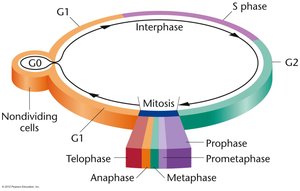
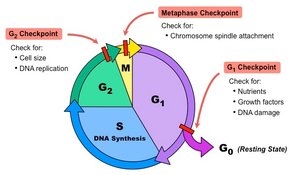
The cell cycle consists of interphase (growth and DNA replication) and the M phase (mitosis or meiosis and cytokinesis). Checkpoints regulate progression and ensure fidelity.
Interphase: G1 (growth), S (DNA synthesis), G2 (preparation for division).
M phase: Mitosis (nuclear division) and cytokinesis (cytoplasmic division).
Checkpoints: G1, G2, and metaphase checkpoints monitor for errors.
Mitosis: Stages and Key Features
Mitosis is the process by which a cell replicates and segregates its chromosomes to produce two genetically identical daughter cells.
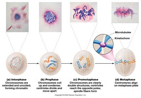
Prophase: Chromosomes condense, centrioles divide, nuclear envelope breaks down.
Prometaphase: Chromosomes move to the metaphase plate, spindle fibers form.
Metaphase: Chromosomes align at the metaphase plate, spindle fibers attach to kinetochores.
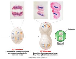
Anaphase: Sister chromatids separate and migrate to opposite poles.
Telophase: Chromosomes arrive at poles, nuclear envelope reforms, cytokinesis occurs.
Chromatin, Centrioles, and Centrosomes
Chromatin is the DNA-protein complex forming chromosomes. Centrioles are structures inside the centrosome, which organize microtubules during cell division.
Centrosome: Cytoplasmic region containing centrioles.
Microtubules: Form the spindle apparatus for chromosome movement.
Chromosome Structure and Centromere Location
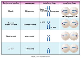
Chromosomes are classified based on centromere position: metacentric, submetacentric, acrocentric, and telocentric.
Centromere location | Designation | Metaphase shape | Anaphase shape |
|---|---|---|---|
Middle | Metacentric | Sister chromatids, centromere in middle | Migration to poles |
Between middle and end | Submetacentric | p arm, q arm | Migration to poles |
Close to end | Acrocentric | Short p arm, long q arm | Migration to poles |
At end | Telocentric | Centromere at end | Migration to poles |
Cohesin and Separase: Chromatid Cohesion and Separation
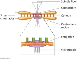
Cohesin is a protein complex holding sister chromatids together. Separase cleaves cohesin, allowing chromatids to separate during anaphase.
Shugoshin: Protects cohesin at centromeres during early stages.
Kinetochore: Protein structure at centromere for spindle attachment.
Genetic Consequences of Mitosis
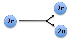
Mitosis produces two cells genetically identical to the parent cell, each with a full complement of chromosomes and approximately half the cytoplasm and organelles.
Counting Chromosomes and DNA Molecules
The number of chromosomes is determined by the number of functional centromeres. The number of DNA molecules doubles after replication but halves after cell division.
Unreplicated chromosome: One DNA molecule per chromosome.
Replicated chromosome: Two DNA molecules per chromosome (sister chromatids).
Meiosis: Mechanism and Consequences
Overview of Meiosis
Meiosis is a specialized cell division producing haploid gametes, reducing chromosome number by half and increasing genetic variation.
Meiosis I: Homologous chromosomes separate, chromosome number is halved.
Meiosis II: Sister chromatids separate, similar to mitosis.
Stages: Each division has prophase, metaphase, anaphase, and telophase.
Genetic Variation in Meiosis
Meiosis results in unique combinations of chromosomes due to independent assortment and crossing-over.
Independent assortment: Random distribution of homologous chromosomes.
Crossing-over: Exchange of genetic material between homologous chromosomes during prophase I.
Formula for combinations: where n is the number of chromosome pairs.
Prophase I Substages
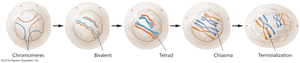
Prophase I is divided into five substages: leptonema, zygonema, pachynema, diplonema, and diakinesis.
Leptonema: Chromomere formation, homology search begins.
Zygonema: Homologs align, synaptonemal complex forms, bivalents visible.
Pachynema: Chromosomes condense, tetrads form.
Diplonema: Chromatids begin to separate, chiasmata visible.
Diakinesis: Nuclear envelope breaks down, spindle fibers attach.
Meiosis I and II: Key Events
Meiosis I separates homologous chromosomes, while meiosis II separates sister chromatids. Each results in haploid cells.
Meiosis I: Reductional division (2n → 1n).
Meiosis II: Equational division (chromatids separate).
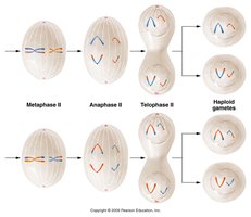
Consequences of Meiosis
Meiosis produces four genetically distinct haploid cells from each diploid cell. Chromosome number is reduced by half, and genetic diversity is increased.
Genetic diversity: Due to crossing-over and independent assortment.
Haploid gametes: Each gamete contains one member of each homologous pair.
Spermatogenesis and Oogenesis
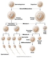
Spermatogenesis produces four haploid spermatids from each primary spermatocyte. Oogenesis produces one ovum and polar bodies, with unequal cytoplasmic division.
Spermatogenesis: Primary spermatocyte → two secondary spermatocytes → four spermatids.
Oogenesis: Primary oocyte → one ovum + polar bodies.
Polar bodies: Small cells with little cytoplasm, useful for genetic diagnostics.
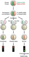
Polar Bodies and Genetic Diagnostics
Polar bodies can be used in IVF to diagnose genetic defects or disease alleles in eggs.
Polar body diagnosis: Identifies eggs without disease alleles or chromosomal abnormalities.
Zona pellucida: Membrane surrounding ovum, retains polar bodies for diagnostics.
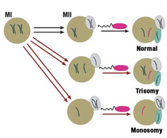
Oogenesis and Chromosomal Aberrations
Aneuploidies, or abnormal chromosome numbers, often result from errors in meiosis, especially in oogenesis. The risk increases with maternal age.
Aneuploidy: Deviations from normal chromosome number (e.g., trisomy, monosomy).
Maternal age effect: Frequency of aneuploidies increases sharply after age 35.
Summary
Chromosomes exist in homologous pairs in diploid organisms.
Mitosis partitions chromosomes into identical diploid cells (2n → 2n).
Meiosis creates haploid gametes and enhances genetic variation (2n → 1n).
Gamete development differs between spermatogenesis and oogenesis.
