 Indietro
Chromosome and DNA Structure: Organization, Visualization, and Compaction in Eukaryotic Cells
Guida di studio - Note intelligenti
 Appunti personalizzati basati sui tuoi materiali, ampliati con definizioni chiave, esempi e contesto.
The Nucleus and Chromosomal Organization
Structure and Function of the Nucleus
The nucleus is a defining organelle of eukaryotic cells, containing the majority of the cell's genetic material. It is surrounded by a dynamic nuclear envelope and contains nuclear pores, chromatin, and a nucleolus. The nucleus is the site of gene expression regulation and houses 23 pairs of chromosomes in humans, totaling approximately 3.2 billion base pairs of DNA.
Nuclear Envelope: Double lipid bilayer that separates nuclear contents from the cytoplasm.
Nuclear Pores: Channels that regulate transport of molecules between the nucleus and cytoplasm.
Nucleolus: Site of ribosomal RNA synthesis and ribosome assembly.
Chromatin: DNA-protein complex that packages DNA within the nucleus.
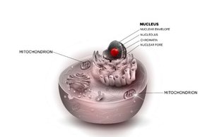
Example: Mature red blood cells lack nuclei, while all other human cells (except platelets) contain nuclei.
Chromosomes and Chromatin
Chromosome Visualization and Chromatin Structure
Chromosomes are only distinctly visible during metaphase of cell division. In non-dividing cells, DNA exists as chromatin, a diffuse, filamentous mass. Chromatin's three-dimensional organization is crucial for gene regulation and genome function.
Metaphase Chromosomes: Highly condensed, visible under a microscope during cell division.
Chromatin: DNA-protein complex, less condensed, present during interphase.
Clinical Application: Chromosome analysis (karyotyping) requires cells to be arrested in metaphase.
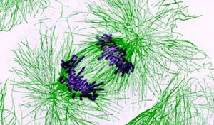
Additional info: Chromatin structure influences accessibility of DNA to transcription machinery and is a focus of epigenetics research.
Karyotypes and Chromosome Classification
Normal Human Karyotype
A karyotype is a visual representation of all chromosomes in a cell, arranged in pairs. Humans have 22 pairs of autosomes and one pair of sex chromosomes (XX for females, XY for males). Chromosomes are numbered by size, with chromosome 1 being the largest.
Autosomes: Chromosomes 1–22, not involved in sex determination.
Sex Chromosomes: X and Y; X is larger and contains more genes, Y is smaller and carries male-determining genes.
Clinical Use: Karyotyping is used to detect chromosomal abnormalities.
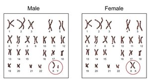
Example: Down syndrome is caused by an extra copy of chromosome 21, visible in a karyotype.
Chromosome Structure and Nomenclature
Basic Chromosome Anatomy
Each chromosome has a characteristic structure with two arms separated by a centromere. The short arm is called the p-arm, and the long arm is the q-arm. Chromosome ends are called telomeres, and the centromere is a region of repetitive DNA important for segregation during cell division.
Centromere: Constriction point, essential for proper chromosome segregation.
Telomere: Repetitive DNA at chromosome ends, protects against degradation.
G-banding: Chromosomes stained with Giemsa dye show characteristic light and dark bands (G-bands), used for identifying chromosomal regions.
Standard Nomenclature: Chromosomal locations are denoted by chromosome number, arm (p or q), and band number (e.g., 15p11). Sub-bands are indicated with decimals (e.g., 15p21.1). Regions near the centromere are labeled 'cen', and ends as 'ter' or 'tel'.
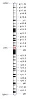
Example: The region 15q11–q13 is associated with Prader-Willi and Angelman syndromes.
Genetic Mapping and Linkage
Centimorgans and Linkage Analysis
Before the Human Genome Project, geneticists estimated distances between genes using linkage analysis. The centimorgan (cM) is a unit of genetic distance based on recombination frequency during meiosis. One centimorgan represents a 1% chance of recombination between two loci.
Linkage: Genes close together on a chromosome tend to be inherited together.
Centimorgan: 1 cM = 1% recombination frequency.
Historical Use: Widely used in classical genetics, especially in model organisms like Drosophila melanogaster (fruit fly).
Additional info: Modern genome mapping uses physical distances (base pairs) rather than recombination units.
DNA Structure and Base Pairing
Chemical Structure of DNA
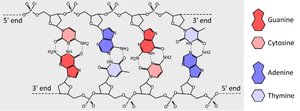
Deoxyribonucleic acid (DNA) is a double-stranded helix composed of a sugar-phosphate backbone and four types of nitrogenous bases: adenine (A), thymine (T), guanine (G), and cytosine (C). The backbone consists of 5-carbon sugars (deoxyribose) linked by phosphate groups.
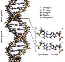
Purines: Adenine and guanine (two-ring structures).
Pyrimidines: Thymine and cytosine (single-ring structures).
Base Pairing: A pairs with T via two hydrogen bonds; G pairs with C via three hydrogen bonds (Watson-Crick base pairing).
Antiparallel Strands: One strand runs 5’ to 3’, the other 3’ to 5’.
Example: High G-C content DNA is more stable due to stronger triple hydrogen bonds.
Double Helix and Molecular Details
The DNA double helix has major and minor grooves, which are important for protein binding and gene regulation. The structure is stabilized by hydrogen bonds between bases and hydrophobic interactions among stacked bases.
Major/Minor Grooves: Allow access for DNA-binding proteins.
Separation: DNA strands can be separated by enzymes (in vivo) or by heat/salt (in vitro).
Additional info: DNA can adopt alternative conformations (A-DNA, Z-DNA) under certain conditions.
Chromatin Structure and DNA Compaction
Nucleosomes and Higher-Order Packaging
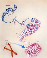
To fit the long DNA molecules into the nucleus, DNA is wrapped around histone proteins to form nucleosomes. Each nucleosome consists of about 147 base pairs of DNA wrapped around a histone octamer. Nucleosomes are connected by linker DNA and further coiled into higher-order structures, ultimately forming metaphase chromosomes.
Nucleosome: "Bead" of DNA wrapped around histone proteins.
Histone Octamer: Core of eight histone proteins.
Chromatin Fiber: Nucleosomes coil into a 30 nm fiber, then loop and condense further.
Example: If stretched linearly, human DNA would be about 2 meters long, but compaction allows it to fit inside a nucleus only a few micrometers in diameter.
Types of Chromatin and Epigenetic Regulation
Chromatin exists in two main forms: euchromatin (less condensed, transcriptionally active) and heterochromatin (more condensed, transcriptionally inactive). Constitutive heterochromatin contains repetitive DNA (e.g., centromeres, telomeres), while facultative heterochromatin can be reversibly silenced or activated via epigenetic modifications such as histone modification.
Histone Modification: Alters chromatin structure and gene accessibility.
Epigenetics: Regulation of gene expression without altering DNA sequence.
Additional info: Chromatin remodeling is essential for processes like cell differentiation and response to environmental signals.
