 Indietro
DNA Structure and the Experimental Evidence for DNA as Genetic Material
Guida di studio - Note intelligenti
 Appunti personalizzati basati sui tuoi materiali, ampliati con definizioni chiave, esempi e contesto.
Chapter 9: DNA Structure and Analysis
Introduction
This chapter explores the chemical structure of DNA, the experimental evidence supporting DNA as the genetic material, and the historical discoveries that shaped our understanding of molecular genetics. Key experiments and concepts are discussed, including the structure of DNA, the nature of its components, and the pivotal studies that established DNA as the hereditary material in living organisms.
DNA Structure
Chemical Bonds and Components of DNA
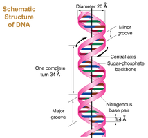
Phosphodiester Bonds: Nucleotides in a DNA strand are linked by phosphodiester bonds, which connect the 5' phosphate group of one nucleotide to the 3' hydroxyl group of the next.
Nitrogenous Bases: DNA contains two types of nitrogenous bases: purines (adenine and guanine) and pyrimidines (cytosine and thymine).
Four Nitrogenous Bases: The four bases in DNA are adenine (A), guanine (G), cytosine (C), and thymine (T).
Base Pairing: Cytosine pairs with guanine via three hydrogen bonds; adenine pairs with thymine via two hydrogen bonds.
Antiparallel Strands: The two DNA strands run in opposite directions (5' to 3' and 3' to 5').
Direction of Synthesis: New DNA strands are synthesized in the 5' to 3' direction during replication.
Deoxyribose Sugar: The sugar in DNA is deoxyribose, a five-carbon sugar lacking an oxygen atom at the 2' position.
Phosphate Groups: Phosphate groups form the backbone of DNA, linking nucleotides and providing structural stability.
Antiparallel Definition: DNA strands are described as antiparallel because one strand runs 5' to 3' and the other 3' to 5', allowing complementary base pairing.
Example: If one DNA strand has the sequence 5’-ACGTT-3’, the complementary strand is 3’-TGCAA-5’.
X-ray Diffraction and the Helical Structure of DNA
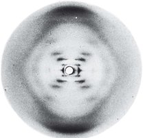
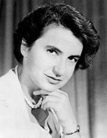
X-ray diffraction studies, notably by Rosalind Franklin, revealed the helical structure of DNA and periodicities characteristic of its double helix.
3.4 Å Periodicity: Indicates the regular spacing of base pairs along the helix.
Helical Structure: The X-ray pattern provided critical evidence for the double helix model.
Historical Discoveries in DNA Research
Key Figures and Milestones
Friedrich Miescher (1869): Discovered nucleic acids in cell nuclei.
Gregor Mendel (1866): Established the basic laws of inheritance.
Chromosome Theory (1902): Linked chromosomes to heredity (Sutton, Boveri, Morgan).
Frederick Griffith (1928): Demonstrated the "transforming principle" in bacteria.
Avery, MacLeod, and McCarty (1944): Identified DNA as the transforming principle.
Hershey and Chase (1952): Confirmed DNA as the genetic material using bacteriophages.
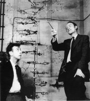
Watson and Crick (1953): Proposed the double helix model of DNA structure.
Modern Advances: Human Genome Project, CRISPR-Cas9 gene editing (Doudna & Charpentier, 2020).
Experimental Evidence for DNA as Genetic Material
Griffith’s Transformation Experiment
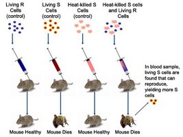
Frederick Griffith's experiments with Streptococcus pneumoniae demonstrated that a "transforming principle" could transfer genetic information from dead virulent bacteria to live non-virulent bacteria, suggesting the existence of a molecular basis for heredity.
Avery, MacLeod, and McCarty’s Experiment
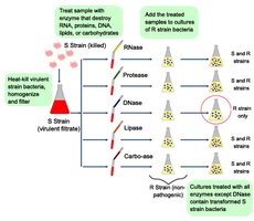
This experiment identified DNA as the "transforming principle" by showing that only DNA, and not proteins or RNA, could transform non-virulent bacteria into virulent forms. Enzymatic destruction of DNA prevented transformation, confirming DNA's role in heredity.
Hershey-Chase Experiment
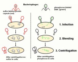
Hershey and Chase used bacteriophages labeled with radioactive isotopes (32P for DNA, 35S for protein) to show that DNA, not protein, enters bacterial cells and directs viral replication. This provided definitive evidence that DNA is the genetic material.
32P labels DNA because phosphorus is present in DNA but not in proteins.
35S labels protein because sulfur is present in certain amino acids but not in DNA.
Limitation of 14C: Carbon-14 is present in both DNA and proteins, so it cannot distinguish between them.
Direct and Indirect Evidence for DNA as Genetic Material
Indirect Evidence
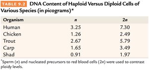
DNA Distribution: The amount of DNA correlates with chromosome number and ploidy in gametes and diploid cells, unlike proteins.
Organelle DNA: Mitochondria and chloroplasts contain their own DNA and perform genetic functions.
Organism | n | 2n |
|---|---|---|
Human | 3.25 | 7.30 |
Chicken | 1.26 | 2.49 |
Trout | 2.67 | 5.79 |
Carp | 1.65 | 3.49 |
Shad | 0.91 | 1.97 |
Direct Evidence
Recombinant DNA Technology: Eukaryotic genes inserted into bacterial DNA can be expressed, as in the production of human insulin.
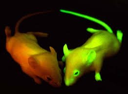
Transgenic Animals: Human genes introduced into animals (e.g., mice) are expressed and inherited, demonstrating the universality of DNA as genetic material.
Checkpoints for Understanding
Recognize the three main structural components of DNA and the concept of 5’ to 3’ polarity.
Understand phosphodiester bonds and the antiparallel double helix structure stabilized by hydrogen bonds (Chargaff’s rules).
Appreciate the similarities and differences between DNA and RNA.
Summarize the direct and indirect evidence supporting DNA as the genetic material in living organisms.
