 Indietro
Mitosis and Meiosis: Cellular Basis of Genetic Inheritance
Guida di studio - Note intelligenti
 Appunti personalizzati basati sui tuoi materiali, ampliati con definizioni chiave, esempi e contesto.
Mitosis and Meiosis
Introduction to Genetic Material and Cell Division
Genetic material in living organisms is composed of nucleic acid DNA, which is organized into chromosomes. In eukaryotes, the transmission of genetic material involves two fundamental processes: mitosis and meiosis. These processes ensure the accurate distribution of genetic information during cellular reproduction and are central to the study of genetics.
Chromosome Theory of Inheritance
Historical Foundations
Mendel’s studies established the principles of inheritance, suggesting that heredity results in discontinuous variation through dominance-recessive relationships.
Nageli and Wiesmann proposed that both parents contribute equally to the traits of offspring.
Boveri and Sutton observed parallels between chromosome behavior during meiosis and Mendelian inheritance patterns.
The chromosome theory of inheritance states that inheritance patterns of traits can be explained by the transmission patterns of chromosomes during meiosis and fertilization.
Chromosome Structure and Organization
General Features of Chromosomes
Chromosomes are structures within cells that contain genetic material (genes).
Composed of DNA (genetic material) and proteins (provide structure).
In eukaryotes, the DNA-protein complex is called chromatin.
Chromosomes in Prokaryotes vs. Eukaryotes
Prokaryotes (e.g., bacteria): Usually have a single, circular chromosome, lack a nucleus, and have no membrane-bound organelles.
Eukaryotes: Have a nucleus with two or more linear chromosomes, and contain membrane-bound organelles such as mitochondria and chloroplasts (which also contain DNA).

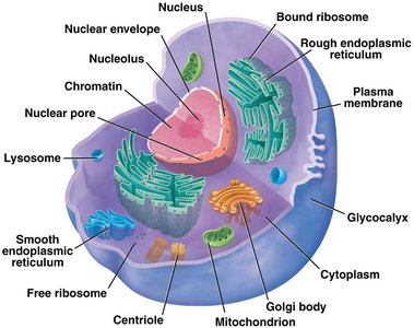
Cytoskeleton and Chromosome Movement
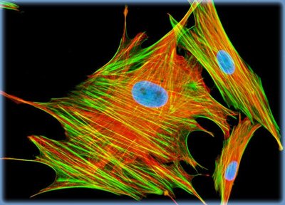
The cytoskeleton, composed of microtubules (tubulin) and microfilaments (actin), provides structural support and is essential for chromosome movement during cell division.
Centrioles and Spindle Formation
Centrioles, located in the centrosome, organize spindle fibers for chromosome movement during mitosis and meiosis.
Centromeres and Chromosome Classification
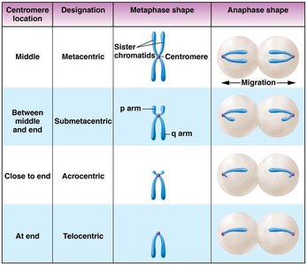
The centromere is a constricted region on the chromosome that determines its shape and is essential for proper segregation during cell division. Chromosomes are classified based on centromere position:
Centromere Location | Designation | Metaphase Shape | Anaphase Shape |
|---|---|---|---|
Middle | Metacentric | Sister chromatids with centromere in the middle | Migration to opposite poles |
Between middle and end | Submetacentric | p arm (short), q arm (long) | Migration to opposite poles |
Close to end | Acrocentric | Centromere near one end | Migration to opposite poles |
At end | Telocentric | Centromere at the end | Migration to opposite poles |
Homologous Chromosomes
Most eukaryotes are diploid, possessing two sets of chromosomes (homologs).
Homologous chromosomes are similar in size, banding pattern, centromere location, and gene content, but may have different alleles.
The physical location of a gene on a chromosome is called its locus.
The Cell Cycle and Mitosis
Phases of the Cell Cycle
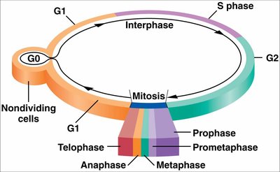
The cell cycle consists of interphase (G1, S, G2) and the M phase (mitosis and cytokinesis). Cells may also enter a non-dividing state called G0.
G1 phase: Cell growth and preparation for DNA replication.
S phase: DNA replication; chromosomes duplicate to form sister chromatids.
G2 phase: Final growth and preparation for mitosis.
M phase: Mitosis (nuclear division) and cytokinesis (cytoplasmic division).
Mitosis: Stages and Mechanisms
Mitosis is subdivided into five phases: prophase, prometaphase, metaphase, anaphase, and telophase. The primary purpose is to distribute replicated chromosomes equally to two daughter cells, ensuring genetic consistency.
Prophase: Chromosomes condense, spindle apparatus forms.
Prometaphase: Nuclear envelope breaks down, spindle fibers attach to kinetochores.
Metaphase: Chromosomes align at the metaphase plate.
Anaphase: Sister chromatids separate and move to opposite poles.
Telophase: Chromosomes decondense, nuclear envelope reforms.
Summary of Mitosis
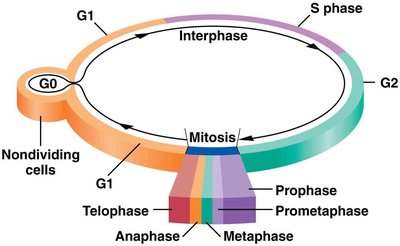
Mitosis produces two genetically identical daughter cells, each with the same chromosome number as the parent cell.
Essential for growth, repair, and asexual reproduction in multicellular organisms.
Meiosis: Basis of Sexual Reproduction
Overview of Meiosis
Meiosis is the process by which haploid gametes are produced from diploid cells. It involves two successive divisions (meiosis I and II), each with prophase, prometaphase, metaphase, anaphase, and telophase stages.
Reduces chromosome number by half (2n → n).
Introduces genetic variation through independent assortment and crossing over.
Gametogenesis
Spermatogenesis: Occurs in testes; produces four haploid sperm cells from one diploid spermatogonial cell.
Oogenesis: Occurs in ovaries; produces one haploid egg and polar bodies from one diploid oogonium due to asymmetric divisions.
Genetic Variation in Meiosis
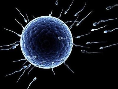
Random segregation of homologous chromosomes and crossing over during meiosis generate genetic diversity among gametes.
Key Terms and Concepts
Diploid (2n): Cell with two sets of chromosomes.
Haploid (n): Cell with one set of chromosomes (e.g., gametes).
Homologous chromosomes: Chromosome pairs with similar structure and gene content.
Sister chromatids: Identical copies of a chromosome, joined at the centromere after DNA replication.
Kinetochore: Protein complex at the centromere where spindle fibers attach during cell division.
Allele: Different versions of a gene found at the same locus on homologous chromosomes.
Summary Table: Mitosis vs. Meiosis
Feature | Mitosis | Meiosis |
|---|---|---|
Number of divisions | 1 | 2 |
Number of daughter cells | 2 | 4 |
Chromosome number in daughter cells | Diploid (2n) | Haploid (n) |
Genetic identity | Identical to parent | Genetically unique |
Role | Growth, repair, asexual reproduction | Sexual reproduction, genetic diversity |
