 Indietro
Study Guide: Mitosis and Meiosis (Genetics Chapter 2)
Guida di studio - Note intelligenti
 Appunti personalizzati basati sui tuoi materiali, ampliati con definizioni chiave, esempi e contesto.
Mitosis and Meiosis: Cellular Basis of Genetic Transmission
Introduction to Chromosomes and Cell Division
Genetic material in living organisms is composed of DNA, organized into chromosomes. In eukaryotes, chromosomes are transmitted via mitosis and meiosis. Mitosis occurs in somatic cells, producing genetically identical cells, while meiosis occurs in sex cells, producing gametes or spores with half the chromosome number.
Cell Structure and Genetic Function
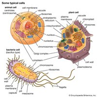
Cell structure is closely tied to genetic function. There are two main cell types:
Prokaryotic cells: Bacteria and archaea, lacking a membrane-bound nucleus.
Eukaryotic cells: Protists, plants, fungi, and animals, with a membrane-bound nucleus.
All cells share common features: plasma membrane, DNA, and ribosomes.
Plasma Membrane and Cell Wall
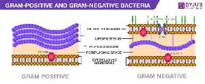
Plasma membrane: Delimits the cell from its environment.
Cell wall: Present in plants (cellulose) and bacteria (peptidoglycan).
Glycocalyx and Receptor Molecules
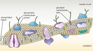
Glycocalyx: Cell coat on animal cells, made of glycoproteins and polysaccharides, provides biochemical identity.
Receptor molecules: Recognition sites for transferring chemical signals across the membrane.
Nucleus and Nucleoid
Nucleus: Membrane-bound, houses DNA in eukaryotes; nucleolus synthesizes rRNA.
Nucleoid: Region in prokaryotes where DNA is located, not membrane-bound.
Cytoplasm and Organelles
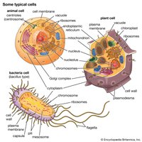
Cytoplasm: Contains organelles and cytosol.
Cytoskeleton: Tubules (microtubules, tubulin) and filaments (microfilaments, actin) provide structure.
Endoplasmic Reticulum (ER): Smooth ER (lipid synthesis), Rough ER (protein synthesis).
Mitochondria: Site of ATP production via respiration.
Chloroplasts: Site of photosynthesis in plants and algae.
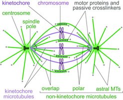
Centrioles: Organize spindle fibers for chromosome movement during cell division.
Chromosomes in Homologous Pairs
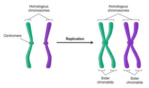
Chromosomes exist in homologous pairs in diploid organisms. Homologous chromosomes carry genes for the same traits but may have different alleles.
Alleles: Alternative forms of the same gene.
Centromere: Constricted region, determines chromosome shape (metacentric, submetacentric, acrocentric, telocentric).
Sister chromatids: Identical copies formed by DNA replication, joined at the centromere.
Diploid and Haploid Chromosome Numbers
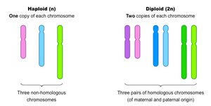
Diploid (2n): Two sets of chromosomes (e.g., humans: 46 chromosomes, 23 pairs).
Haploid (n): One set of chromosomes (gametes).
Karyotype
Karyotype: Physical appearance and arrangement of chromosomes in a cell.
Autosomes: Non-sex chromosomes.
Sex chromosomes: X and Y, non-homologous in humans.
Mitosis: Partitioning Chromosomes
Mitosis is the process by which chromosomes are partitioned into two genetically identical daughter cells. It is followed by cytokinesis, which divides the cytoplasm.
Cell Cycle
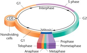
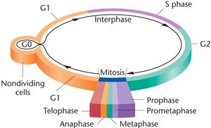
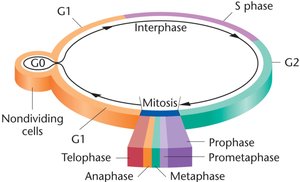
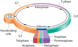
Interphase: Includes G1 (growth), S (DNA synthesis), G2 (preparation for mitosis), and G0 (nondividing state).
Mitosis: Prophase, Prometaphase, Metaphase, Anaphase, Telophase.
Stages of Mitosis
Prophase: Chromosomes condense, nuclear envelope breaks down, centrioles move to poles.
Prometaphase: Chromosomes move to metaphase plate, spindle fibers form.
Metaphase: Chromosomes align at metaphase plate, spindle fibers attach to kinetochores.
Anaphase: Sister chromatids separate and move to opposite poles.
Telophase: Chromosomes uncoil, nuclear envelope reforms, spindle fibers disappear.
Cytokinesis: Division of cytoplasm, forming two new cells.
Cell Cycle Regulation and Checkpoints
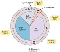
Kinases and cyclins: Regulate cell cycle progression.
Checkpoints: Monitor for errors during cell division.
Meiosis: Creation of Haploid Gametes and Genetic Variation
Meiosis reduces the chromosome number by half, producing haploid gametes or spores. It consists of two divisions: Meiosis I (reductional) and Meiosis II (equational).
Meiosis I: Prophase I Substages
Leptonema: Chromosomes begin to condense.
Zygonema: Synapsis begins, homologous chromosomes pair.
Pachynema: Crossing over occurs between nonsister chromatids.
Diplonema: Chiasmata visible, synapsis ends.
Diakinesis: Nuclear membrane disintegrates, chromosomes prepare for metaphase.
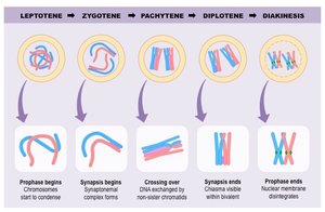
Meiosis I: Metaphase I, Anaphase I, Telophase I
Metaphase I: Tetrads align at metaphase plate.
Anaphase I: Homologous chromosomes separate (nondisjunction may occur).
Telophase I: Nuclear membrane reforms, cytokinesis produces two haploid cells.
Meiosis II: Prophase II, Metaphase II, Anaphase II, Telophase II
Prophase II: Chromosomes condense again.
Metaphase II: Chromosomes align at metaphase plate.
Anaphase II: Sister chromatids separate.
Telophase II: Four haploid cells are produced.
Genetic Variation
Crossing over: Exchange of genetic material between homologous chromosomes during prophase I increases genetic diversity.
Biparental inheritance: Each diploid organism receives alleles from both parents.
Development of Gametes: Spermatogenesis vs. Oogenesis
Gamete development differs between males and females:
Spermatogenesis: Primary spermatocyte undergoes meiosis to produce four haploid spermatids.
Oogenesis: Primary oocyte undergoes meiosis, but only one daughter cell receives most cytoplasm and matures into an ovum; the others become polar bodies.
Physical Nature of Chromosomes: Electron Microscopy
Chromosomes are visible only during mitosis and meiosis. Chromatin fibers coil and condense to form chromosomes, as revealed by electron microscopy.
Summary Table: Comparison of Mitosis and Meiosis
Feature | Mitosis | Meiosis |
|---|---|---|
Number of divisions | 1 | 2 |
Chromosome number in daughter cells | Diploid (2n) | Haploid (n) |
Genetic variation | None (identical cells) | High (crossing over, independent assortment) |
Function | Growth, repair | Gamete/spore production |
Key Terms and Concepts
Chromatin: Diffuse network of DNA during nondivisional phases.
Centromere: Region where sister chromatids are joined.
Kinetochore: Protein complex at centromere, attaches to spindle fibers.
Cohesin: Protein complex holding sister chromatids together.
Separase: Enzyme that degrades cohesin.
Shugoshin: Protects cohesin from separase.
Chiasma: Site of crossing over in meiosis.
Tetrad: Four chromatids formed during meiosis I.
Equations
Diploid number: Haploid number: DNA replication:
