 Indietro
Biological Membranes and Membrane Transport: Structure, Function, and Mechanisms
Guida di studio - Note intelligenti
 Appunti personalizzati basati sui tuoi materiali, ampliati con definizioni chiave, esempi e contesto.
The Structure and Function of Biological Membranes
Biological Membranes: Composition and Organization
Biological membranes are essential structures that define cell boundaries and regulate the movement of substances into and out of cells. The primary component of these membranes is the phospholipid bilayer, which is amphipathic—containing both hydrophilic and hydrophobic regions. Embedded within this bilayer are other molecules such as proteins and cholesterol, which contribute to membrane function and fluidity. This arrangement is described by the Fluid Mosaic Model, which states that membranes are dynamic and composed of a mosaic of proteins that float in or on the fluid lipid bilayer.
Phospholipids: Major structural component, forming a bilayer.
Proteins: Integral and peripheral proteins serve various functions.
Cholesterol: Modulates membrane fluidity and stability.
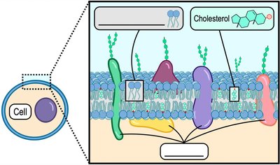
Example: The major components of membranes are phospholipids, proteins, and cholesterol.
Types of Membrane Proteins
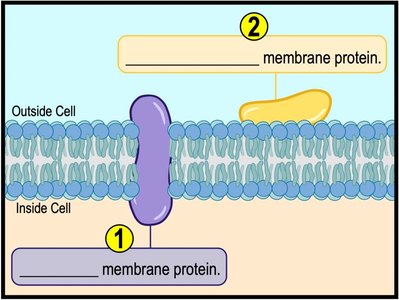
Membrane-associated proteins are classified based on their location and interaction with the membrane:
Integral Membrane Proteins: Span the entire bilayer and are embedded within the membrane.
Peripheral Membrane Proteins: Located on the surface or perimeter of the membrane, not embedded.
Functions of Membrane Proteins
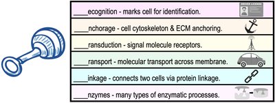
Membrane proteins perform a wide variety of functions, including:
Recognition: Mark cells for identification.
Anchorage: Anchor the cell cytoskeleton and extracellular matrix.
Transduction: Act as signal molecule receptors.
Transport: Facilitate molecular transport across the membrane.
Linkage: Connect two cells via protein linkage.
Enzymes: Catalyze many types of enzymatic processes.
Concentration Gradients and Diffusion
Concentration Gradients
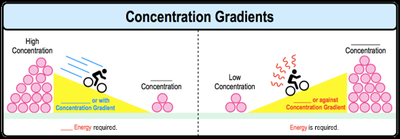
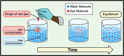
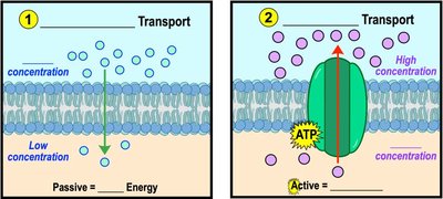
A concentration gradient refers to the difference in the concentration of a substance between two areas. Molecules tend to move down their concentration gradient (from high to low concentration), a process that does not require energy. Movement up the gradient (from low to high concentration) requires energy input.
Diffusion
Diffusion is the movement of a substance from an area of higher concentration to an area of lower concentration. This process is driven by the natural tendency of molecules to spread out and does not require cellular energy.
Example: Diffusion of a dye in water demonstrates how molecules move until equilibrium is reached.
Membrane Transport: Selective Permeability
Selective Permeability of Biological Membranes
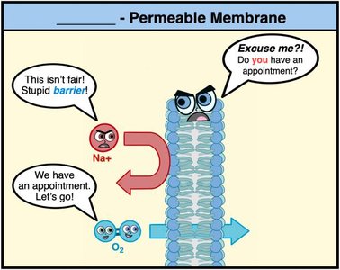
Biological membranes are selectively permeable, meaning they regulate what substances can cross. This property allows cells to maintain internal conditions distinct from their external environment.
Molecules That Freely Cross Membranes
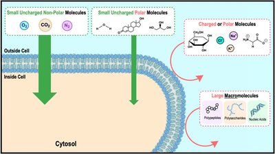
Some molecules can diffuse across the membrane without the aid of proteins:
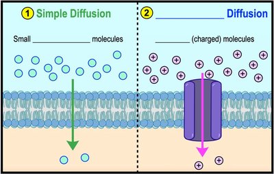
Small, uncharged, nonpolar molecules (e.g., O2, CO2)
Large, charged, or polar molecules require protein facilitation.

Types of Membrane Transport
Overview of Membrane Transport
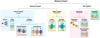
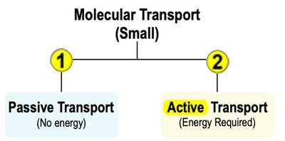
Membrane transport is categorized into molecular transport (for small molecules) and bulk transport (for large molecules). Molecular transport can be passive (no energy required) or active (energy required).
Passive vs. Active Transport
There are two general types of molecular transport:
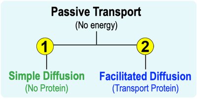
Passive Transport: Moves molecules from high to low concentration without energy input.
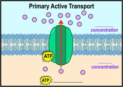
Active Transport: Moves molecules from low to high concentration, requiring energy (usually ATP).
Classes of Membrane Transport Proteins
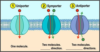
Transport proteins are classified by their mechanism:
Uniporters: Transport one molecule at a time in one direction.
Symporters: Cotransport two or more molecules in the same direction.
Antiporters: Cotransport two or more molecules in opposite directions.
Osmosis and Tonicity
Osmosis
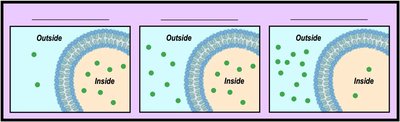
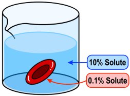
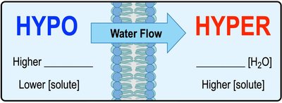
Osmosis is the passive diffusion of water across a semi-permeable membrane. The direction of water flow depends on the tonicity—the relative concentration of solutes in the solutions.
Hypotonic: Lower solute concentration outside the cell.
Isotonic: Equal solute concentrations inside and outside.
Hypertonic: Higher solute concentration outside the cell.
Direction of Osmosis
Water moves from hypotonic to hypertonic solutions if solutes cannot diffuse across the membrane. Water always moves from areas of higher water concentration to lower water concentration.
Environmental Tonicity Effects on Cells
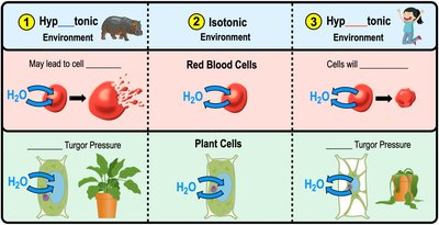
The tonicity of the environment affects cell shape and function:
Hypotonic: Water enters cells, causing swelling and possible lysis. Preferred by plant cells for turgor pressure.
Isotonic: Water enters and exits at equal rates; preferred by animal cells.
Hypertonic: Water exits cells, causing dehydration and shriveling.
Simple and Facilitated Diffusion
Types of Passive Transport
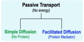
Passive transport includes simple diffusion and facilitated diffusion:
Simple Diffusion: Direct diffusion of small, uncharged molecules through the membrane.
Facilitated Diffusion: Diffusion of charged or larger molecules facilitated by transport proteins.
Active Transport
Primary and Secondary Active Transport
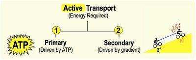
Active transport requires energy to move molecules against their concentration gradient:
Primary Active Transport: Directly driven by ATP hydrolysis.
Secondary Active Transport: Driven by the concentration gradient established by primary active transport.
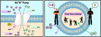
Na+/K+ Pump (Primary Active Transport Example)
The sodium-potassium pump is an antiporter that moves Na+ and K+ ions in opposite directions, using energy from ATP hydrolysis. It exports three Na+ ions and imports two K+ ions per cycle.
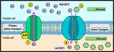
Secondary Active Transport: Sodium-Glucose Cotransporter
Secondary active transport uses the Na+ gradient established by primary active transport to power the transport of glucose against its gradient.
Endocytosis and Exocytosis
Bulk Transport: Endocytosis and Exocytosis
Large biomolecules are transported across cell membranes via endocytosis and exocytosis:
Endocytosis: Macromolecule engulfment by the cell membrane, allowing entry into the cell via a lipid vesicle.
Exocytosis: Vesicle fusion with the cell membrane, allowing contents to exit the cell.
Types of Endocytosis:
Phagocytosis: Large, solid material is taken in (cell "eating").
Pinocytosis: Small, liquid material is taken in (cell "drinking").
Receptor-Mediated Endocytosis: Specific form using receptor proteins.
Example: White blood cells engulf bacteria using phagocytosis.
Exocytosis: Hormones, neurotransmitters, and digestive enzymes are secreted out of the cell by exocytosis.
*Additional info: The notes cover all major aspects of membrane structure and transport, including passive and active mechanisms, osmosis, and bulk transport, making them highly relevant for Human Biology students studying cell structure and function.*
