 Indietro
Structure, Function, and Life Cycles of Sponges and Cnidarians
Guida di studio - Note intelligenti
 Appunti personalizzati basati sui tuoi materiali, ampliati con definizioni chiave, esempi e contesto.
Sponges: Structure and Function
Basic Anatomy of Sponges
Sponges (phylum Porifera) are simple, multicellular animals that lack true tissues and organs. Their bodies are adapted for filter feeding, allowing water to flow through specialized structures.
Spongocoel: The central cavity where water collects before exiting.
Pores: Small openings in the sponge's body wall through which water enters.
Epidermis: The outer cell layer providing protection.
Osculum: The large opening at the top where water exits.
Choanocytes: Flagellated cells that create water currents and trap food particles.
Amoebocytes: Cells that distribute nutrients and produce spicules for structural support.
Mesohyl: Gelatinous matrix within the sponge containing cells and spicules.
Spicules: Structural elements made of silica or calcium carbonate.
Example: Sponges filter water to obtain food, with choanocytes capturing particles and amoebocytes distributing nutrients. 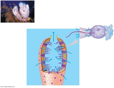
Cnidarians: Structure and Life Cycle
Body Forms and Anatomy
Cnidarians (phylum Cnidaria) are aquatic animals with specialized stinging cells called cnidocytes. They exhibit two main body forms: polyp and medusa.
Polyp: Cylindrical, sessile form with tentacles surrounding the mouth.
Medusa: Free-swimming, bell-shaped form with tentacles hanging down.
Gastrovascular cavity: Central digestive compartment.
Epidermis: Outer tissue layer.
Gastrodermis: Inner tissue layer lining the cavity.
Mesoglea: Gel-like layer between epidermis and gastrodermis.
Example: Jellyfish (medusa) and hydra (polyp) are classic cnidarian forms. 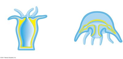
Cnidocyte and Nematocyst Function
Cnidocytes are specialized cells unique to cnidarians, used for defense and prey capture.
Nematocyst: A capsule within the cnidocyte containing a coiled thread that can discharge rapidly.
Trigger: A hair-like structure that, when touched, causes the nematocyst to fire.
Function: The discharged thread can inject toxins into prey or predators.
Example: Sea anemones and jellyfish use nematocysts to immobilize prey. 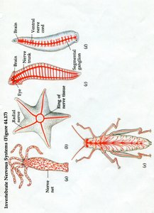
Life Cycle of Cnidarians
Cnidarians often alternate between polyp and medusa forms in their life cycle, involving both sexual and asexual reproduction.
Asexual reproduction: Budding in polyps produces new individuals.
Sexual reproduction: Medusae release gametes; fertilization produces a zygote.
Planula: Free-swimming larval stage that settles to form a new polyp.
Example: The life cycle of a typical jellyfish includes both polyp and medusa stages. 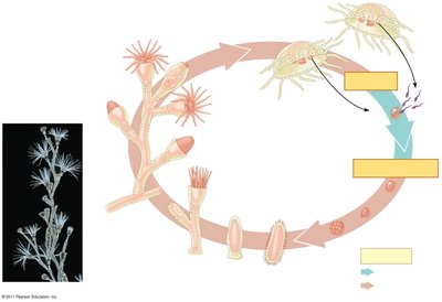
Classification and Diversity of Cnidarians
Major Classes of Cnidarians
Cnidarians are classified into several classes based on their body forms and life cycles.
Hydrozoa: Mostly colonial polyps, some with medusa stages.
Scyphozoa: True jellyfish, dominant medusa stage.
Anthozoa: Sea anemones and corals, only polyp stage.
Example: Hydra (Hydrozoa), Aurelia (Scyphozoa), and corals (Anthozoa). 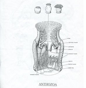
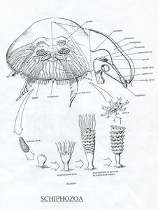
Additional Structures and Movements
Locomotion in Cnidarians
Some cnidarians, such as hydra, exhibit unique movements like somersaulting to change location.
Somersaulting: Hydra attaches with tentacles, flips its body, and reattaches with its basal disc.
Example: Hydra moves by somersaulting, a simple form of locomotion. 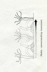
Summary Table: Comparison of Sponge and Cnidarian Structure
Feature | Sponges | Cnidarians |
|---|---|---|
Body Symmetry | Asymmetrical | Radial |
Tissues | No true tissues | True tissues |
Digestive System | Intracellular (choanocytes) | Gastrovascular cavity |
Reproduction | Asexual and sexual | Asexual (budding) and sexual |
Specialized Cells | Choanocytes, amoebocytes | Cnidocytes (nematocysts) |
Key Terms and Definitions
Spongocoel: Central cavity in sponges.
Osculum: Large opening for water exit in sponges.
Choanocyte: Flagellated cell for water movement and food capture in sponges.
Polyp: Sessile body form in cnidarians.
Medusa: Free-swimming body form in cnidarians.
Cnidocyte: Stinging cell in cnidarians.
Nematocyst: Capsule in cnidocyte for prey capture.
Planula: Larval stage in cnidarians.
Equations and Formulas
Surface Area to Volume Ratio
The efficiency of diffusion in simple animals like sponges and cnidarians depends on their surface area to volume ratio.
Formula:
Application: High surface area to volume ratio facilitates efficient nutrient and gas exchange.
Conclusion
Sponges and cnidarians represent early evolutionary stages of animal life, with unique adaptations for feeding, defense, and reproduction. Understanding their structure and life cycles provides insight into the diversity and complexity of multicellular organisms.
