 Indietro
Chapter 3: The Integumentary System – Medical Terminology Study Guide
Guida di studio - Note intelligenti
 Appunti personalizzati basati sui tuoi materiali, ampliati con definizioni chiave, esempi e contesto.
Integumentary System Overview
Functions of the Integumentary System
The integumentary system is essential for protecting the body and maintaining homeostasis. It consists of the skin and its accessory organs, performing several vital functions:
Protection: Acts as a two-way barrier, preventing pathogens and harmful substances from entering, and keeping critical body fluids inside.
Temperature Regulation: Controls heat loss and retention through sweat production and blood vessel dilation/constriction.
Sensory Reception: Contains receptors for temperature, pain, touch, and pressure.
Secretion: Produces sweat and sebum, which lubricate and cool the skin.
Organs: Skin, hair, nails, sebaceous glands, and sweat glands.
Anatomy and Physiology of the Skin
Layers of the Skin
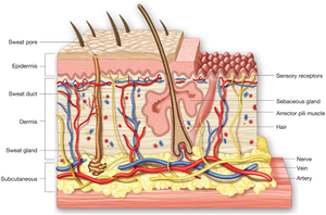
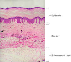
The skin is the largest organ in the body, composed of three main layers:
Epidermis: The thin, outer membrane layer made of stratified squamous epithelium. Contains the basal layer, where cells grow and produce keratin, and melanocytes that produce melanin for UV protection.
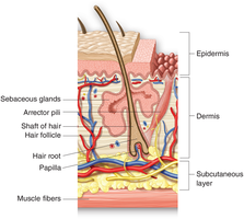
Dermis: The middle, fibrous connective tissue layer. Contains blood vessels, lymph vessels, sensory receptors, hair follicles, sweat and sebaceous glands, and muscle fibers. Provides strength and houses accessory structures.
Subcutaneous Layer (Hypodermis): The deepest layer, composed of fat cells (lipocytes). Provides insulation and protects deeper tissues.
Epidermis
Stratified Squamous Epithelium: Flat, scale-like cells arranged in strata.
Basal Layer: Deepest layer; cells grow and push old cells upward, which die and fill with keratin, forming a waterproof barrier.
Melanocytes: Produce melanin, giving skin its color and protecting against UV rays.
Dermis
Corium: Living tissue with a good blood supply.
Collagen Fibers: Provide strength and elasticity.
Structures: Hair follicles, sweat and sebaceous glands, blood and lymph vessels, sensory receptors, nerve fibers, and muscle fibers.
Fingerprints: Dermal ridges create unique patterns.
Subcutaneous Layer
Hypodermis: Composed of lipocytes (fat cells).
Functions: Insulates the body and protects deeper tissues from injury.
Accessory Organs of the Skin
Hair
Hair is an accessory organ consisting of the follicle, root, shaft, and arrector pili muscle. It grows as keratinized cells are pushed upward by new cells. Melanin gives hair its color, and the arrector pili muscle contracts to make the hair stand up.
Nails
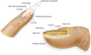
Nails are composed of a flat plate of keratin called the nail body, connected to the underlying tissue by the nail bed. The lunula is at the base, and nails grow from the nail root, covered by the cuticle. The free edge is the exposed part that must be trimmed.
Sebaceous Glands
Function: Release sebum directly into hair follicles, lubricating hair and skin.
Clinical Note: Increased secretion during adolescence can lead to acne; decreased secretion with age contributes to dry skin and wrinkles.
Sweat Glands
Sweat (Sudoriferous) Glands: Coiled glands in the dermis; sweat travels to the surface via ducts and pores.
Function: Sweat cools the body as it evaporates; most sweat is colorless and odorless, but apocrine glands produce body odor.
Medical Terminology: Key Terms
Combining Forms, Prefixes, and Suffixes
Medical terms for the integumentary system are constructed using combining forms, prefixes, and suffixes. Understanding these components is essential for accurate communication and documentation.
Combining Forms: Root words with a vowel, used to connect with other word parts (e.g., derm/o for skin).
Prefixes: Added to the beginning of terms to modify meaning (e.g., epi- meaning above).
Suffixes: Added to the end of terms to indicate procedures, conditions, or diseases (e.g., -itis meaning inflammation).
Example: The term dermatitis combines dermat/o (skin) and -itis (inflammation) to mean inflammation of the skin.
Pathology of the Skin
Burns: Degrees and Assessment
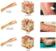
Burns are classified by depth and severity. The three degrees of burns are:
First-degree (Superficial): Affects only the epidermis; causes redness and pain.
Second-degree (Partial thickness): Involves epidermis and part of the dermis; causes blisters and more severe pain.
Third-degree (Full thickness): Extends through the dermis and may affect subcutaneous tissue; causes charring and loss of sensation.
Rule of Nines
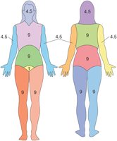
The Rule of Nines is a method for estimating the percentage of body surface area affected by burns. The body is divided into regions, each representing a percentage of the total surface area.
Skin Cancer
Basal Cell Carcinoma: Most common type; rarely metastasizes.
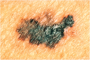
Malignant Melanoma: Highly aggressive; characterized by dark, irregular lesions.
Infectious and Other Skin Disorders
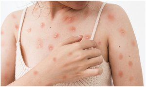
Monkeypox: Viral infection causing characteristic rash.
Clinical Procedures and Pharmacology
Diagnostic and Therapeutic Procedures
Biopsy: Removal of skin tissue for examination.
Skin Grafting: Transplantation of skin to cover damaged areas.
Plastic Surgery: Procedures to repair or reconstruct skin and underlying tissues.
Medications
Antibiotics: Treat bacterial infections.
Antifungals: Treat fungal infections.
Antivirals: Treat viral infections.
Topical Steroids: Reduce inflammation.
Abbreviations
Common abbreviations used in the integumentary system include:
Bx: Biopsy
Derm: Dermatology
SubQ: Subcutaneous
SC: Subcutaneous
Summary Table: Layers and Functions of the Skin
Layer | Main Components | Functions |
|---|---|---|
Epidermis | Stratified squamous epithelium, basal layer, melanocytes | Protection, waterproof barrier, UV protection |
Dermis | Connective tissue, collagen, blood vessels, glands, follicles | Strength, elasticity, houses accessory organs |
Subcutaneous Layer | Lipocytes (fat cells) | Insulation, protection |
Key Equations
Burn surface area estimation (Rule of Nines):
Additional info: Academic context was added to clarify terminology, pathology, and clinical procedures for completeness and exam preparation.
