 Indietro
Digestive System: Medical Terminology and Anatomy for Health Care Professionals
Guida di studio - Note intelligenti
 Appunti personalizzati basati sui tuoi materiali, ampliati con definizioni chiave, esempi e contesto.
Digestive System Overview
Introduction to the Digestive System
The digestive system, also known as the gastrointestinal (GI) tract, is a continuous tube that processes food, absorbs nutrients, and eliminates waste. It includes both primary and accessory organs, each with specialized functions essential for human health.
Primary organs: Mouth, pharynx, esophagus, stomach, small intestine, large intestine
Accessory organs: Salivary glands, liver, gallbladder, pancreas
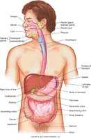
Length: Approximately 30 feet in adults
Main functions:
Digestion: Breakdown of food into absorbable components
Absorption: Uptake of nutrients into blood or lymph
Elimination: Removal of solid waste from the body
Anatomy of the Digestive System
Mouth (Oral Cavity)
The mouth is the entry point for food and is responsible for the initial mechanical and chemical breakdown of food. It is formed by the hard and soft palates, cheeks, tongue, and lips. The vestibule is the space between the cheeks and teeth, and the gingiva (gums) surround the necks of the teeth. The lingual frenulum connects the tongue to the floor of the mouth.
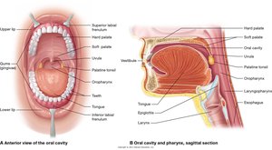
Tongue: Made of skeletal muscle, divided into root, body, and tip; contains taste buds and papillae
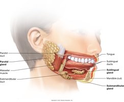
Salivary glands: Three pairs (parotid, submandibular, sublingual) secrete saliva to moisten food and begin digestion
Digestion begins in the mouth: Food is broken apart, moistened, and formed into a bolus
Teeth
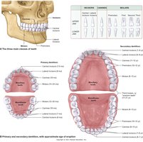
Humans have two sets of teeth: deciduous (baby) and permanent (adult). Teeth are specialized for cutting, tearing, and grinding food.
Deciduous teeth: 20 total (8 incisors, 4 canines, 8 molars)
Permanent teeth: 32 total (8 incisors, 4 canines, 8 premolars, 12 molars)
Types of teeth:
Incisors: Sharp cutting edge
Canines: Pointed for tearing
Premolars: Grinding surface
Molars: Largest, for grinding and pounding
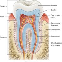
Tooth structure:
Crown: Projects above the gum
Root: Embedded in a bony socket (alveolus)
Neck: Between crown and root
Pulp cavity: Contains dental pulp, vessels, and nerves
Dentin: Main solid portion
Enamel: Covers crown
Cementum: Covers root, anchors periodontal ligament
Pharynx
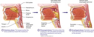
The pharynx (throat) is a muscular tube that serves as a common passageway for food and air. It is divided into three regions: nasopharynx, oropharynx, and laryngopharynx. The epiglottis prevents food from entering the airway during swallowing.
Nasopharynx: Above the soft palate
Oropharynx: Between palate and hyoid bone
Laryngopharynx: Between hyoid bone and entrance to larynx/esophagus
Esophagus
The esophagus is a muscular tube that transports food from the pharynx to the stomach. The lower esophageal sphincter (LES) regulates passage and prevents reflux. Food is moved by peristalsis, a series of coordinated muscular contractions.
Stomach
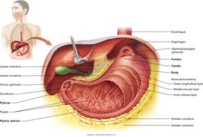
The stomach is a muscular, saclike organ that continues the digestive process. It is divided into the fundus (upper), body (main), and antrum (lower). Rugae are folds that allow expansion and contain glands for digestive juices. Food is mixed and reduced to chyme, which passes through the pyloric sphincter into the small intestine.
Small Intestine
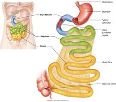
The small intestine is the primary site for digestion and absorption. It is divided into three sections: duodenum, jejunum, and ileum. Villi increase the surface area for absorption, and digestive juices from the liver and pancreas aid in nutrient breakdown.
Duodenum: First 12 inches
Jejunum: Next 8 feet
Ileum: Last 12 feet
Large Intestine
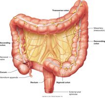
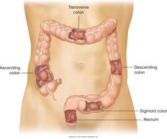
The large intestine absorbs water and forms feces. It consists of the cecum, colon, rectum, and anal canal. The colon is further divided into ascending, transverse, descending, and sigmoid sections.
Accessory Organs of Digestion
Salivary Glands
Salivary glands secrete saliva, which contains enzymes that initiate carbohydrate digestion and lubricate food for swallowing. The three major pairs are the parotid, submandibular, and sublingual glands.
Liver
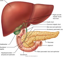
The liver is the largest glandular organ and plays a central role in metabolism, detoxification, and synthesis of important proteins. It produces bile for fat emulsification, stores glycogen, and synthesizes clotting factors.
Gallbladder
The gallbladder stores and concentrates bile produced by the liver. Bile is released into the small intestine to aid in fat digestion.
Pancreas
The pancreas secretes digestive enzymes (pancreatic juice) into the small intestine and produces hormones such as insulin and glucagon for blood sugar regulation.
Medical Terminology: Building Your Vocabulary
Common Terms and Word Parts
cholecystectomy: Surgical removal of the gallbladder (chol/e = bile, cyst = bladder, -ectomy = excision)
diarrhea: Abnormal frequency and liquidity of fecal discharge (dia- = through, -rrhea = flow)
glossectomy: Excision of the tongue (gloss = tongue, -ectomy = excision)
rectocele: Hernia of the rectum (rect/o = rectum, -cele = hernia)
Drug Highlights
Medications Related to the Digestive System
Antacids: Neutralize stomach acid (HCl)
Histamine H2-receptor antagonists: Inhibit gastric acid secretion
Proton-pump inhibitors (PPIs): Suppress gastric acid secretion
Laxatives: Relieve constipation
Antidiarrheal agents: Treat diarrhea
Antiemetics: Prevent or stop vomiting
Emetics: Induce vomiting
Diagnostic and Laboratory Tests
Common Tests for Digestive Disorders
Alcohol toxicology: Measures blood alcohol levels
Carcinoembryonic antigen (CEA): Monitors cancer therapy
Cholangiography: X-ray of bile ducts after dye ingestion
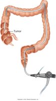
Colonoscopy: Visual examination of the colon with a colonoscope
Comprehensive metabolic panel (CMP): Blood tests for metabolic function
Endoscopic retrograde cholangiopancreatography (ERCP): X-ray of biliary and pancreatic ducts
Esophagogastroduodenoscopy: Endoscopic exam of esophagus, stomach, and small intestine
Gastric analysis: Tests stomach contents for acid, blood, bacteria, bile, and fatty acids
Hepatitis panel: Blood test for hepatitis infection markers
Liver biopsy: Microscopic exam of liver tissue
Occult blood test: Detects hidden blood in stool
Ova and parasites (O&P): Stool test for parasites
Stool culture: Identifies bacteria in stool
Ultrasonography: Visualizes gallbladder or liver using sound waves
Upper GI endoscopy: Visual exam of gastric mucosa
Selected Pathologies and Procedures
Appendectomy and Appendicitis
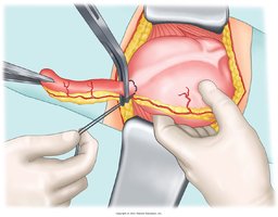
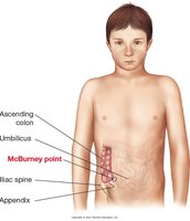
An appendectomy is the surgical removal of the appendix, often performed to treat appendicitis, which is inflammation of the appendix.
Cirrhosis
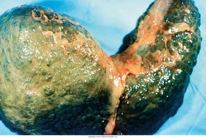
Cirrhosis is a chronic liver disease characterized by scarring and impaired liver function. Common causes include chronic alcohol use and hepatitis.
Colorectal Cancer
Colorectal cancer affects the colon and rectum. Early detection through colonoscopy is critical for effective treatment.
Diverticulitis
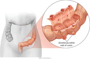
Diverticulitis is inflammation of diverticula (small pouches) in the colon wall, which can cause pain and digestive disturbances.
Gastroesophageal Reflux Disease (GERD)
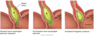
GERD is a condition where stomach acid frequently flows back into the esophagus, causing irritation. It is often due to dysfunction of the lower esophageal sphincter.
Hemorrhoids
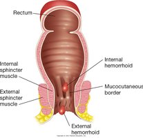
Hemorrhoids are swollen veins in the lower rectum or anus, classified as internal or external. They can cause pain, bleeding, and discomfort.
Peptic Ulcer Disease
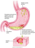
Peptic ulcers are sores that develop on the lining of the stomach, duodenum, or esophagus due to the action of gastric acids.
Volvulus
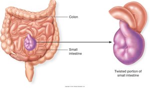
Volvulus is a twisting of the intestine that can cause obstruction and compromise blood flow, requiring prompt medical intervention.
Summary Table: Digestive System Organs and Functions
Organ | Main Function |
|---|---|
Mouth | Mechanical and chemical digestion; forms bolus |
Pharynx | Passageway for food and air; swallowing |
Esophagus | Transports food to stomach via peristalsis |
Stomach | Mixes and digests food; forms chyme |
Small Intestine | Digestion and absorption of nutrients |
Large Intestine | Absorbs water; forms and eliminates feces |
Salivary Glands | Secrete saliva to moisten food and begin digestion |
Liver | Produces bile; metabolism; detoxification |
Gallbladder | Stores and concentrates bile |
Pancreas | Secretes digestive enzymes and hormones |
Key Abbreviations
GI: Gastrointestinal
LES: Lower esophageal sphincter
ERCP: Endoscopic retrograde cholangiopancreatography
CMP: Comprehensive metabolic panel
CEA: Carcinoembryonic antigen
