 Indietro
Gas Exchange and Respiratory System: Medical Terminology Study Guide
Guida di studio - Note intelligenti
 Appunti personalizzati basati sui tuoi materiali, ampliati con definizioni chiave, esempi e contesto.
Gas Exchange in the Respiratory System
Introduction to Gas Exchange
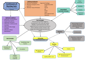
Gas exchange is a fundamental physiological process in which oxygen (O2) is inhaled from the environment, transferred to the bloodstream, and exchanged for carbon dioxide (CO2) in the lungs. This process is essential for cellular metabolism and the removal of metabolic waste.
Definition: Gas exchange refers to the movement of O2 into the blood and CO2 out of the blood, primarily occurring in the alveoli of the lungs.
Location: Alveoli, tiny air sacs surrounded by capillaries, are the primary site of gas exchange.
Transport: Red blood cells (erythrocytes) and hemoglobin are required to transport oxygen throughout the body.
Purpose: To supply tissues with oxygen and remove carbon dioxide.
Anatomy of the Respiratory System
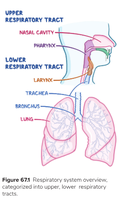
The respiratory system is divided into upper and lower tracts, each with specific structures and functions.
Upper Respiratory Tract: Includes the nasal cavity, pharynx, and larynx.
Lower Respiratory Tract: Includes the trachea, bronchi, bronchioles, and lungs.
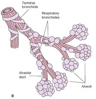
Alveoli: Terminal structures where gas exchange occurs.
Pulmonary Circulation
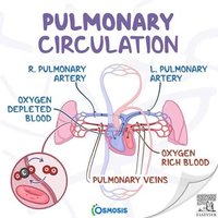
Pulmonary circulation is the movement of blood between the heart and lungs, facilitating gas exchange.
Oxygen-depleted blood is pumped from the right ventricle to the lungs via the pulmonary arteries.
Oxygen-rich blood returns to the left atrium via the pulmonary veins.
Purpose: To replenish oxygen and remove carbon dioxide from the blood.
Mechanisms of Breathing
Breathing involves ventilation, respiration, and perfusion.
Ventilation: The movement of air in and out of the lungs.
Respiration: The exchange of gases at the alveolar level; normal rate is 12-20 breaths per minute.
Perfusion: The flow of blood through the pulmonary capillaries.
Assessment of Gas Exchange
Clinical assessment is crucial for evaluating respiratory function and gas exchange.
Skin color: Cyanosis may indicate hypoxemia.
Respiratory depth and effort: Observing for labored or shallow breathing.
Respiratory rate: Normal range is 12-20 breaths per minute.
Oxygen saturation: Measured by pulse oximetry; normal is 95-100%.
Arterial blood gases (ABGs): Laboratory test to assess O2 and CO2 levels.
Maintaining Patent Airway
A patent airway is essential for effective gas exchange.
Definition: An airway is patent when there is an open pathway between the lungs and the outside world.
Obstruction: Blockage may be partial or total, preventing air from entering the lungs.
Abnormal Lung Sounds
Auscultation of lung sounds helps identify respiratory disorders.
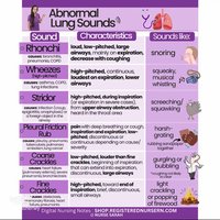
Rhonchi: Low-pitched, snoring sounds; often clear with coughing.
Wheezes: High-pitched, musical sounds; indicate airway narrowing.
Stridor: High-pitched, during inspiration; suggests upper airway obstruction.
Crackles: Bubbling or popping sounds; associated with fluid in alveoli.
Signs and Symptoms of Altered Gas Exchange
Hypoxemia: Low oxygen in the blood; symptoms include shortness of breath (SOB), altered mental status (AMS), confusion, tachycardia, skin/lip discoloration, dizziness.
Hypoxia: Low oxygen in tissues/organs.
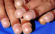
Clubbing: Enlargement of fingertips due to chronic hypoxia.
Respiratory Patterns
Tachypnea: Rapid breathing.
Bradypnea: Slow breathing.
Hypoventilation: Decreased rate or depth of breathing.
Hyperventilation: Increased rate or depth of breathing.
Apnea: Absence of breathing.
Dyspnea: Difficulty breathing.
Orthopnea: Difficulty breathing when lying flat.
Diagnostic Evaluation
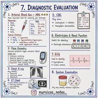
Common Diagnostic Tests
Diagnostic tests are used to assess respiratory function and identify underlying disorders.
Arterial Blood Gas (ABG): Measures O2, CO2, and pH in blood.
Pulse Oximetry: Noninvasive measurement of oxygen saturation.
Chest X-ray: Visualizes lung structure and pathology.
Bronchoscopy: Direct visualization of airways.
Pulmonary angiogram: Assesses blood flow in pulmonary vessels.
Pulmonary V-Q scan: Evaluates ventilation and perfusion.
Thoracentesis: Removal of fluid from pleural space.
Interventions for Gas Exchange
Independent Nursing Interventions
Elevate head of bed: Improves lung expansion.
Maintain airway patency: Suctioning, positioning, and airway management.
Health promotion: Smoking cessation, vaccines, environmental control.
Collaborative/Interprofessional Interventions
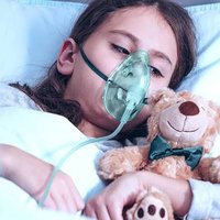
Oxygen administration: Supplemental O2 via various devices.
Pharmacologic therapy: Bronchodilators, corticosteroids, anticholinergics.
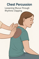
Clearing pulmonary secretions: Chest physiotherapy, fluids, humidification, nebulization.
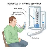
Incentive spirometry: Encourages deep breathing to prevent atelectasis.
Oxygen Delivery Systems
Oxygen can be delivered using several devices, each suited to different clinical needs.
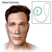
Nasal cannula: Delivers low-flow oxygen; requires patent airway.
Simple mask: Provides moderate oxygen concentration.
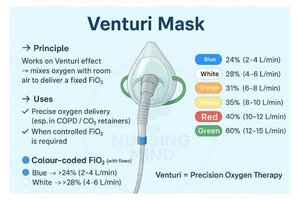
Venturi mask: Delivers precise oxygen concentration; useful in COPD.
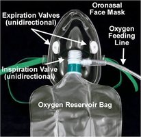
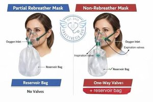
Nonrebreather mask: Delivers high concentration; requires one-way valves and reservoir bag.
Respiratory Disorders
Chronic Obstructive Pulmonary Disease (COPD)
COPD is a group of progressive lung diseases characterized by airflow limitation.
Emphysema: Destruction and enlargement of alveoli, loss of lung elasticity, airway inflammation, fibrosis, narrowing of airways.
Expected findings: SOB, cough, decreased breath sounds.
Asthma
Asthma is a chronic airway disease marked by inflammation and hyperreactivity.
Triggers: Irritants, allergens, pollutants, cold air.
Expected findings: Wheezing, SOB, chest tightness.
Pneumonia
Pneumonia is an infection of the lung parenchyma, caused by bacteria or viruses.
Transmission: Droplets or aerosols inhaled by the host.
Expected findings: SOB, confusion, lethargy, loss of appetite, cough.
Case Study Example
Respiratory Distress in Older Adults
Subjective information: Patient may report SOB, confusion, fatigue, chest discomfort.
Objective findings: Crackles, diminished breath sounds, abnormal vital signs, low oxygen saturation.
Summary Table: Oxygen Delivery Devices
Device | Oxygen Concentration | Clinical Use |
|---|---|---|
Nasal Cannula | 24-40% | Low-flow, mild hypoxemia |
Simple Mask | 40-60% | Moderate O2 needs |
Venturi Mask | 24-60% | Precise O2 delivery, COPD |
Nonrebreather Mask | 60-100% | Severe hypoxemia |
Key Terms and Definitions
Alveoli: Tiny air sacs in the lungs where gas exchange occurs.
Hemoglobin: Protein in red blood cells that binds oxygen.
Hypoxemia: Low oxygen in the blood.
Hypoxia: Low oxygen in tissues.
Pulse Oximetry: Noninvasive measurement of oxygen saturation.
Bronchodilator: Medication that relaxes airway muscles.
Relevant Equations
Oxygen Saturation Calculation:
Minute Ventilation:
Health Promotion and Prevention
Smoking cessation is critical for preventing respiratory diseases.
Vaccination against influenza and pneumonia reduces risk of infection.
Environmental control minimizes exposure to pollutants and allergens.
Summary
Understanding gas exchange and the respiratory system is essential for medical terminology students. Mastery of anatomy, physiology, assessment, interventions, and terminology supports effective clinical practice and patient care. Additional info: Academic context was added to clarify mechanisms, interventions, and diagnostic tests.
