 Indietro
Med Terms Chap 3
Guida di studio - Note intelligenti
 Appunti personalizzati basati sui tuoi materiali, ampliati con definizioni chiave, esempi e contesto.
Dermatology and Medical Terminology
Definition and Word Parts
Dermatology is the medical specialty focused on the study of the skin and its diseases. Understanding medical terminology is essential for proficiency in this area.
Dermat/o-: means skin
-logy: means study of

Anatomy of the Integumentary System
Overview and Major Structures
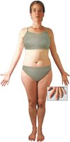
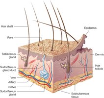
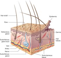
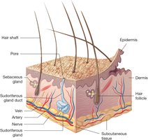
The integumentary system covers most of the body and consists of the skin, nails, and subcutaneous tissue. The skin is the largest organ and is composed of two main layers: the epidermis and dermis.
Epidermis: Thin, outermost layer made of epithelial tissue
Dermis: Thick layer beneath the epidermis, made of connective tissue
Subcutaneous tissue: Connective tissue beneath the dermis, contains adipose (fat) tissue
Word Parts in Anatomy
Medical terms often use word parts to describe anatomical structures:
integument/o-: skin
-ary: pertaining to
cutane/o-: skin
-ous: pertaining to
Epidermis Structure
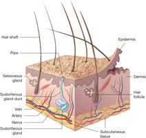
The epidermis consists of two main layers:
Squamous layer: Upper part, contains living and dead cells. Dead cells are filled with keratin, forming a protective, slightly acidic layer that discourages microorganism growth.
Basal layer: Deep part, made of living cells that constantly divide and move upward. The epidermis contains no blood vessels and relies on the dermis for nutrients.
Melanocytes and Skin Pigmentation
Melanocytes are cells in the epidermis that produce melanin, the pigment responsible for skin color and protection against UV light.
Everyone has the same number of melanocytes, but melanin production varies.
Sun exposure increases melanin production; insufficient absorption leads to sunburn.
Dermis Structure
The dermis is composed of connective tissue and contains:
Collagen: Firm, white protein providing strength
Elastin: Elastic, yellow protein providing flexibility
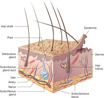
Blood vessels, nerves, hair follicles, sebaceous glands, and sweat glands
Skin and Subcutaneous Tissue
The skin consists of the epidermis and dermis, while the subcutaneous tissue lies beneath and is composed of adipose tissue, providing cushioning and insulation.
Common Diseases and Conditions
Skin Edema
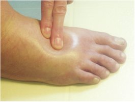
Edema is swelling caused by excess fluid in the tissues. Pitting edema occurs when fingertip pressure leaves a deep indentation.
Types of Skin Lesions
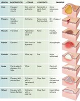
Skin lesions are classified by their appearance, color, and contents.
Lesion | Description | Color | Contents | Example |
|---|---|---|---|---|
Cyst | Elevated, encapsulated | Skin color or erythema | Semisolid or partly fluid | Acne sebaceous cyst |
Fissure | Small, crack-like | Erythema | None | Dry chapped skin |
Macule | Flat circle | Pigmented or erythema | None | Freckle, age spot |
Papule | Elevated | Skin color or erythema | None | Acne whitehead |
Pustule | Elevated | White top | Pus | Acne pimple |
Scale | Flat to slightly elevated | White or silver | None | Dandruff, psoriasis |
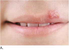
Vesicle | Elevated with pointed top | Erythema | Clear fluid | Herpes, chickenpox |
Wheal | Elevated with broad, flat top | Pale | Clear fluid | Insect bites, urticaria |
Skin Color Conditions
Abnormal skin color can indicate underlying diseases:
Albinism: Genetic condition with lack of melanin, resulting in pale skin, hair, and eyes
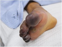
Cyanosis: Bluish discoloration due to lack of oxygen
Necrosis: Tissue death from lack of blood supply, often leading to gangrene
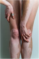
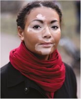
Vitiligo: Autoimmune disorder causing depigmentation in patches


Skin Injuries
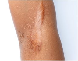
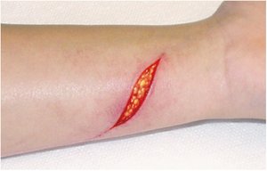
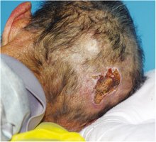
Skin injuries include burns, lacerations, pressure injuries, and abnormal scarring.
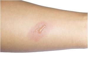
Partial-thickness burn: Separation of epidermis from dermis, forming blisters
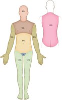
Rule of Nines: Tool for estimating burn surface area
Keloid: Excessive scar tissue formation
Laceration: Deep cut exposing subcutaneous tissue
Pressure injury: Tissue damage from prolonged pressure
Skin Infections and Infestations
Common skin infections include viral, fungal, and allergic conditions.
Herpes simplex: Vesicles on lips (cold sores)
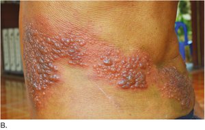
Shingles: Vesicles and crusts along a dermatome
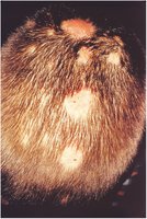
Tinea capitis: Fungal infection of the scalp, also known as ringworm
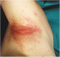
Contact dermatitis: Allergic reaction causing erythema and irritation
Benign and Malignant Neoplasms
Skin neoplasms can be benign or malignant:
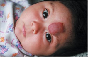
Hemangioma: Benign lesion with dilated blood vessels
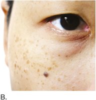
Nevus: Pigmented mole, can be flat or raised
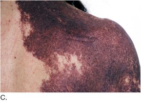
Port-wine stain: Vascular nevus resembling spilled wine
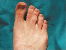
Syndactyly: Fusion of skin and soft tissues between digits
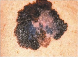
Malignant melanoma: Cancerous lesion with asymmetry, irregular edges, and color variation
Autoimmune Disorders
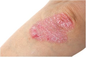
Psoriasis is a chronic autoimmune disorder characterized by red, scaly patches, commonly on elbows and knees.
Anatomy and Physiology of Skin Glands
Sebaceous Glands
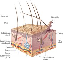
Sebaceous glands are exocrine glands that produce sebum, an oily substance that moisturizes the skin and hair.
Diseases of Sebaceous Glands
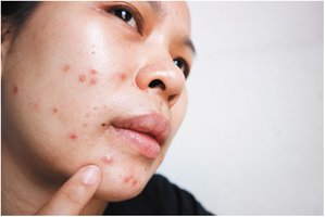
Acne vulgaris: Triggered by increased sebum production during puberty, resulting in papules and pustules
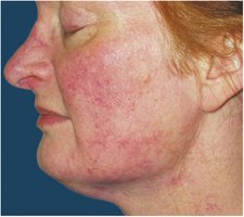
Rosacea: Chronic condition with erythema and dilated blood vessels
Sudoriferous (Sweat) Glands
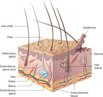
Sudoriferous glands secrete sweat, which is odorless until it contacts skin bacteria. Sweat helps regulate body temperature.
Anatomy of Hair and Nails
Hair Structure and Function
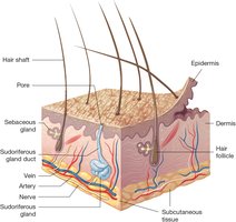
Hair covers most of the body, with color determined by melanocytes. Hair forms in follicles in the dermis and is strengthened by keratin.
Piloerection
Piloerection occurs when the skin is cold, causing hair to stand up and form goosebumps due to contraction of tiny muscles at the base of hair follicles.
Nail Anatomy
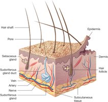
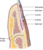
Nails consist of five parts: nail plate, nail bed, lunula, cuticle, and nail root. Nails protect the fingertips and aid in fine motor tasks.
Diseases of the Nail
Onychomycosis: Fungal infection causing discoloration, thickening, and deformation of nails
Subcutaneous Tissue
The subcutaneous tissue is loose connective tissue beneath the dermis, composed of adipose cells (lipocytes). It cushions, insulates, and protects internal organs.
Physiology of the Integumentary System
Protection
The skin acts as the body's first line of defense against injury and infection. Keratin makes the skin waterproof, and sweat/sebum contain antibodies and enzymes that kill bacteria.
Repair
When injured, the skin repairs itself through cell migration and division. Deep wounds form blood clots and scabs, with new cells filling the wound.
Sensation
Sensory receptors in the dermis detect touch, pressure, vibration, pain, and temperature, sending signals to the nervous system for interpretation.
Vitamin D Synthesis
UV rays convert epidermal cholesterol into vitamin D, which is essential for calcium absorption and bone health. Sun exposure requirements vary by skin tone.
Thermoregulation
Thermoregulation involves controlling body temperature:
Heating: Subcutaneous tissue stores fat, piloerector muscles contract
Cooling: Sudoriferous glands produce sweat, dermal blood vessels dilate
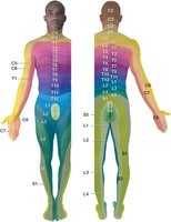
Dermatomes
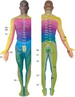
A dermatome is an area of skin that sends sensory information to a specific spinal nerve. Each dermatome is named according to the spinal cord level it connects to (C, T, L, S).
Homeostasis
Homeostasis is the process of maintaining balance and stability in body systems. The integumentary system plays a key role in thermoregulation and signaling other medical conditions.
Laboratory, Diagnostic, and Medical Procedures
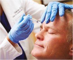
Botox Injection
Botox is a diluted neurotoxin used for cosmetic and medical purposes, such as reducing wrinkles.
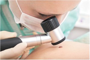
Skin Examination
Dermatologists use magnifying lenses to examine skin lesions, which may require biopsy for diagnosis.
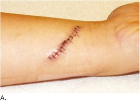
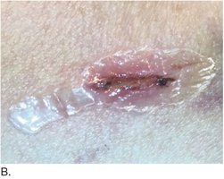
Wound Closure
Wounds may be closed with sutures or surgical glue. Sutures can be placed in layers, with deeper ones absorbed by the body.
Drugs: Routes of Administration
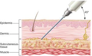
Hypodermic injection delivers drugs beneath the skin, into the subcutaneous tissue.
Surgical Procedures
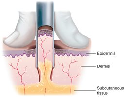
Punch biopsy: Uses a circular cutter to remove a core of skin tissue for examination
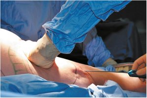
Liposuction: Removes fat from subcutaneous tissue for cosmetic purposes
Abbreviations
Common Abbreviations in Dermatology
Medical terminology in dermatology often uses abbreviations for efficiency. Examples include "sub Q" for subcutaneous tissue.
