 Indietro
Introduction to Imaging and Radiologic Sciences: Medical Terminology and Applications
Guida di studio - Note intelligenti
 Appunti personalizzati basati sui tuoi materiali, ampliati con definizioni chiave, esempi e contesto.
Introduction to Medical Radiation Sciences
Overview of Medical Radiation Sciences
Medical radiation sciences involve the use of various forms of energy to create images of the human body for diagnostic and therapeutic purposes. These energies are carefully controlled to ensure patient safety while providing valuable information for medical care.
Medical Imaging: The process of creating visual representations of the interior of a body for clinical analysis and medical intervention.
Ionizing vs. Non-Ionizing Energy: Some imaging modalities use ionizing radiation (e.g., X-rays), which can alter atomic structure and potentially harm tissue, while others use non-ionizing energy (e.g., ultrasound, MRI), which does not have this effect.
Energy Forms Used in Medical Imaging
Types of Energy and Their Applications
Different imaging modalities utilize specific types of energy, each with unique properties and clinical uses.
Electromagnetic Energy: Includes X-rays (ionizing), radio waves (non-ionizing), and gamma rays (ionizing).
Sound Energy: Used in medical sonography (ultrasound), which is non-ionizing and safe for various applications, including obstetrics.
Electrical Energy: Used in electrocardiography (ECG) to record the electrical activity of the heart.
Heat (Thermal) Energy: Used in thermography to detect heat patterns and blood flow in body tissues.
Magnetic Energy: Utilized in magnetic resonance imaging (MRI) to produce detailed images of organs and tissues.
Nuclear Energy: Involves gamma radiation and radioactive isotopes in nuclear medicine for functional imaging.
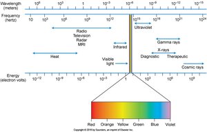
Figure: The electromagnetic spectrum illustrates the range of energies used in medical imaging, from radio waves to gamma rays. Placement on the spectrum is determined by frequency and wavelength.
Major Imaging Modalities
Radiography (X-ray Imaging)
Radiography uses X-rays, a form of ionizing electromagnetic energy, to create images of the body's internal structures. It is one of the most common and essential diagnostic tools in medicine.
Ionization: X-rays can ionize atoms, which is the basis for image formation but also requires careful safety protocols to minimize tissue damage.
Applications: Used for imaging bones, detecting fractures, and diagnosing various diseases.
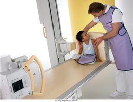
Example: A radiologic technologist positions a patient for an X-ray examination, ensuring proper technique and safety.
Diagnostic Medical Sonography (Ultrasound)
Medical sonography uses high-frequency sound waves to produce images of organs and tissues. It is a non-ionizing modality, making it safe for repeated use and for sensitive populations such as pregnant women.
Applications: Commonly used in obstetrics, cardiology, and abdominal imaging.
Credentialing: Requires formal education and certification.
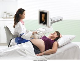
Example: Sonography is widely used for fetal imaging during pregnancy.
Magnetic Resonance Imaging (MRI)
MRI uses strong magnetic fields and radio waves to generate detailed images of the body's internal structures. It is a non-ionizing imaging technique, making it safe for most patients.
Applications: Particularly useful for imaging soft tissues such as the brain, muscles, and joints.
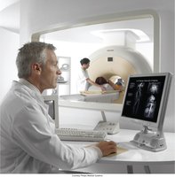
Computed Tomography (CT) Scanning
CT scanning employs X-ray energy and advanced computer algorithms to create cross-sectional images of the body. It provides detailed views of internal anatomy and is essential for diagnosing a wide range of conditions.
Technique: The X-ray tube and detectors rotate around the patient, acquiring multiple images that are reconstructed into slices.
Training: CT technologists require specialized education.
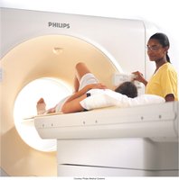
Nuclear Medicine
Nuclear medicine uses radioactive isotopes (radiopharmaceuticals) that emit gamma radiation to visualize physiological processes in the body. It is particularly valuable for assessing organ function and detecting abnormalities at the molecular level.
Gamma Radiation: The primary energy form used in nuclear medicine imaging.
Applications: Includes bone scans, cardiac stress tests, and cancer detection.
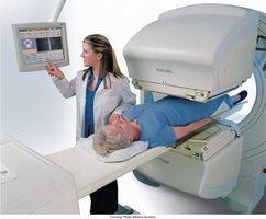
Cardiovascular Interventional Technology (CVIT)
CVIT utilizes X-rays and contrast materials to visualize blood vessels and heart anatomy. Procedures are performed in specialized angiography labs and often involve catheterization.
Applications: Includes angioplasty, stent placement, and cardiac catheterization.
Teamwork: Technologists work closely with angiographers and other specialists.
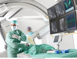
Radiation Therapy
Radiation therapy uses high-energy ionizing radiation to treat malignant tumors. It is a cornerstone of cancer treatment and requires precise planning and delivery to maximize tumor control while minimizing damage to healthy tissue.
Oncology Team: Radiation therapists collaborate with medical dosimetrists and oncologists to develop and administer treatment plans.
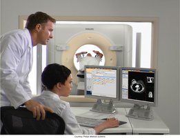
History of Medicine and Radiology
Milestones in Medical History
The history of medicine spans over 5000 years, with significant advancements shaping modern healthcare.
Hippocrates: Known as the "Father of Western Medicine."
Scientific Method: Applied to medicine in the 17th century, revolutionizing medical research and practice.
Louis Pasteur: Discovered the role of germs in disease processes.
DNA: The human genetic code was discovered in the mid-20th century, opening new avenues for diagnosis and treatment.
Discovery of X-rays and the Birth of Radiology
Radiology began with the discovery of X-rays by Wilhelm Conrad Röntgen on November 8, 1895. This breakthrough revolutionized medical diagnostics and earned Röntgen the Nobel Prize in Physics in 1901.
First X-ray Image: The first radiograph was of Röntgen’s wife’s hand, marking the beginning of diagnostic imaging.
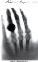
Example: The first X-ray image ever produced, showing the bones of a hand and a ring.
Radiologic Sciences as a Career
Career Opportunities and Specialties
The field of radiologic sciences offers a wide range of career paths, often starting with general radiography and expanding into specialized areas through additional education and certification.
Specialties: Radiography, MRI, CT, CVIT, medical sonography, nuclear medicine, radiation therapy, mammography, bone densitometry, education, administration, and roles in commercial firms.
Career Ladder: Opportunities for advancement exist in clinical practice, education, research, and administration.
Radiology in the Health Care Team
Roles and Responsibilities
Radiologic technologists are essential members of the healthcare team, working alongside physicians, nurses, and allied health professionals to provide diagnostic and therapeutic services.
Team Members: Include medical doctors (MD), doctors of osteopathy (DO), nurses, allied health personnel, and non-clinical support staff.
Allied Health: Most health careers outside of medicine and nursing are considered allied health professions.
Conclusion
X-rays, discovered by W.C. Röntgen in 1895, laid the foundation for modern medical imaging. Radiologic sciences encompass a variety of diagnostic and therapeutic modalities, all of which play a critical role in patient care. The field offers diverse career opportunities and requires a commitment to safety, education, and professional growth.
