 Indietro
Med Terms Chap 4
Guida di studio - Note intelligenti
 Appunti personalizzati basati sui tuoi materiali, ampliati con definizioni chiave, esempi e contesto.
Gastroenterology: Introduction and Terminology
Definition and Medical Language
Gastroenterology is the medical specialty focused on the anatomy, physiology, diseases, and treatment of the gastrointestinal (GI) system. Understanding medical terminology is essential for describing the structures, functions, and disorders of the GI tract.
Gastr/o-: Refers to the stomach.
Enter/o-: Refers to the intestine.
-logy: Means the study of.
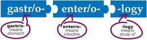
Example: Gastroenterology literally means "the study of the stomach and intestines."
Overview of the Gastrointestinal System
Major Structures and Pathway
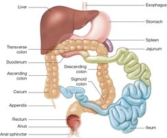
The GI system is a continuous tube with associated glands and organs. It is responsible for digestion, absorption of nutrients, and elimination of waste. The pathway begins at the mouth and ends at the anus.
Oral cavity (mouth)
Pharynx (throat)
Esophagus
Stomach
Small intestine (duodenum, jejunum, ileum)
Large intestine (cecum, colon, rectum, anus)
Accessory organs: liver, gallbladder, pancreas
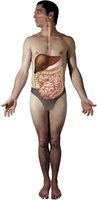
Anatomy: Upper Gastrointestinal System
Oral Cavity and Pharynx
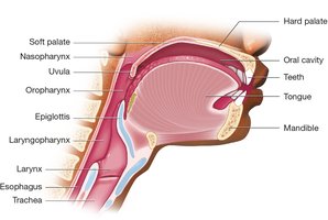
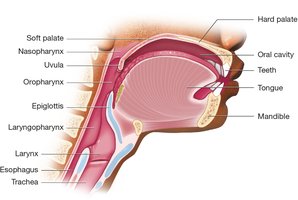
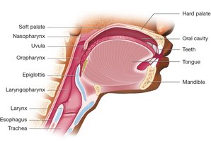
The oral cavity contains the teeth, gums, tongue, and the hard and soft palates. Food passes from the oral cavity into the pharynx (throat) and then the esophagus.
Teeth: Responsible for mastication (chewing).
Tongue: Assists in mixing food and swallowing; contains taste receptors.
Hard and soft palates: Form the roof of the mouth; the soft palate and uvula help direct food into the pharynx.
Salivary Glands
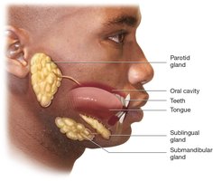
Salivary glands secrete saliva, which contains enzymes that begin the process of digestion and lubricate food for swallowing.
Parotid glands: Located anterior to the ears.
Sublingual glands: Located under the tongue.
Submandibular glands: Located beneath the mandible (lower jaw).
Pharynx and Larynx
The pharynx is a shared passageway for food and air, divided into the nasopharynx, oropharynx, and laryngopharynx. The larynx, located just below the pharynx, remains open during breathing and closes during swallowing to prevent aspiration.
Epiglottis: A flap that covers the larynx during swallowing, directing food into the esophagus.
Esophagus
The esophagus is a muscular tube that connects the pharynx to the stomach. Peristalsis, a series of coordinated muscle contractions, moves food toward the stomach.
Stomach
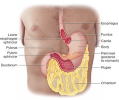
The stomach is a muscular organ divided into four regions: cardia, fundus, body, and pylorus. It mixes food with gastric juices to form chyme and regulates its passage into the small intestine via sphincters.
Lower esophageal sphincter: Prevents reflux of stomach contents.
Pyloric sphincter: Controls the release of chyme into the duodenum.
Omentum: Fatty membrane protecting abdominal organs.
Diseases of the Upper GI Tract
Dyspepsia
Dyspepsia refers to indigestion, often caused by excess stomach acid, reflux, overeating, spicy foods, or stress.

Esophageal Varices
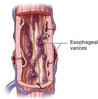
Esophageal varices are dilated veins in the esophageal mucosa, which can bleed if irritated. They are often associated with liver disease and portal hypertension.
Gastric Ulcer
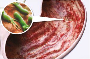
A gastric ulcer is an erosion of the stomach lining, often caused by Helicobacter pylori infection or chronic use of NSAIDs. It can lead to bleeding and pain.
Diagnostic and Medical Procedures: Upper GI
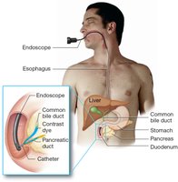
Endoscopic Retrograde Cholangiopancreatography (ERCP)
ERCP is a procedure in which an endoscope is passed through the mouth to the duodenum, allowing visualization and injection of contrast dye to image the bile and pancreatic ducts.
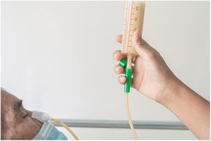
Nasogastric (NG) Tube
An NG tube is inserted through the nose into the stomach for feeding or medication administration when oral intake is not possible.
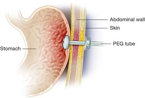
Percutaneous Endoscopic Gastrostomy (PEG) Tube
A PEG tube is a permanent feeding tube inserted through the abdominal wall into the stomach for long-term nutritional support.
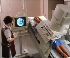
Upper Gastrointestinal Series
This radiologic procedure involves swallowing barium contrast to visualize the upper GI tract on x-ray.
Anatomy: Lower Gastrointestinal System
Small Intestine
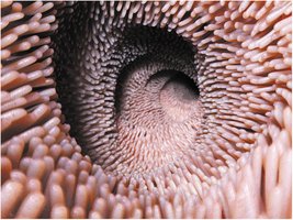
The small intestine is a long, narrow tube divided into three segments: duodenum, jejunum, and ileum. It is lined with villi to maximize nutrient absorption.
Duodenum: First segment, about 10 inches long.
Jejunum: Middle segment, about 8 feet long.
Ileum: Last segment, about 12 feet long.
Large Intestine
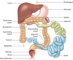
The large intestine absorbs water and forms feces. It consists of the cecum (with appendix), colon (ascending, transverse, descending, sigmoid), rectum, and anus. Haustra are pouches that expand to accommodate waste.
Cecum and Ileocecal Valve
The cecum is a short sac at the beginning of the large intestine. The ileocecal valve prevents backflow of waste into the small intestine.
Colon
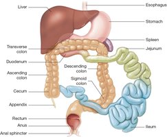
The colon is the largest part of the large intestine and is divided into four regions: ascending, transverse, descending, and sigmoid colon. Haustra are visible as puckered pouches.
Diseases of the Lower GI Tract
Intussusception and Volvulus
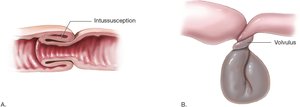
Intussusception occurs when one part of the intestine slides into another, while volvulus is a twisting of the intestine. Both conditions can obstruct blood flow and peristalsis.
Diverticula and Diverticulitis
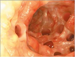
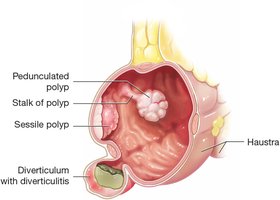
Diverticula are pouches in the colon wall where feces can become trapped. Infection of a diverticulum is called diverticulitis.
Crohn Disease
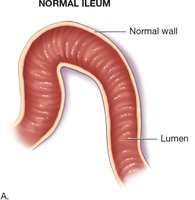
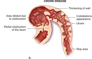
Crohn disease is a chronic inflammatory condition of the GI tract, often affecting the ileum and colon. It causes wall thickening, ulcers, and partial obstruction.
Colonic Polyps
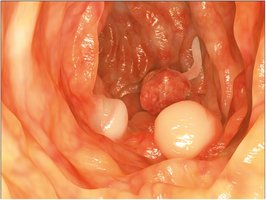
Polyps are abnormal growths in the colon mucosa. Sessile polyps are flat, while pedunculated polyps are attached by a stalk. Some polyps can become cancerous.
Colostomy and Stoma
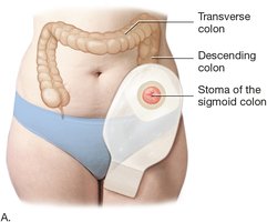
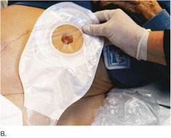
A colostomy is a surgical procedure creating an opening (stoma) from the colon to the abdominal wall for fecal diversion. A colostomy bag collects waste.
Colonoscopy
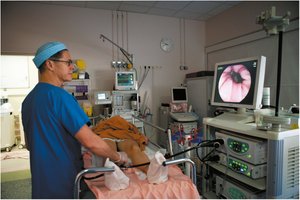
A colonoscope is inserted through the anus to visualize the rectum and colon for diagnostic and therapeutic purposes.
Barium Enema
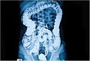
Barium contrast is inserted through the rectum to visualize the colon on x-ray, highlighting the haustra and any abnormalities.
Diseases: Abdominal Wall and Abdominopelvic Cavity
Hernia
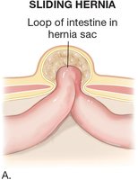
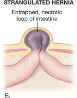
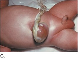
A hernia occurs when an organ or tissue protrudes through a weak spot in the abdominal wall. Types include sliding and strangulated hernias.
Sliding hernia: Intestine moves in and out of the hernia sac.
Strangulated hernia: Intestine is trapped, leading to tissue death.
Omphalocele: Congenital hernia at the umbilicus.
Peritonitis
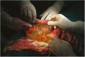
Peritonitis is inflammation of the peritoneum, often due to perforation of the GI tract and leakage of contents into the abdominal cavity.
Anatomy: Accessory Organs
Liver
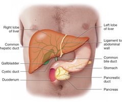
The liver is the largest solid organ, producing bile for fat digestion. Bile flows through the biliary tree to the gallbladder and duodenum.
Hepatocytes: Liver cells that produce bile.
Biliary tree: Network of bile ducts.
Gallbladder
The gallbladder stores and concentrates bile, releasing it into the duodenum in response to fatty chyme.
Pancreas
The pancreas secretes digestive enzymes (amylase, lipase, proteases) into the duodenum via the pancreatic duct.
Diseases: Liver, Gallbladder, and Pancreas
Liver Disease
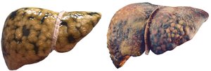
Liver diseases include fatty liver (steatosis), cirrhosis (scarring and nodules), and cancer. Cirrhosis impairs liver function and can lead to complications such as portal hypertension and ascites.
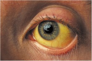
Jaundice
Jaundice is yellow discoloration of the skin and sclera due to elevated bilirubin, often from liver dysfunction or bile duct obstruction.
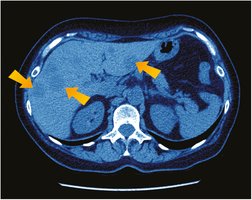
Liver Cancer
Liver cancer appears as masses or nodules on imaging studies such as CT scans.
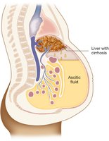
Ascites
Ascites is the accumulation of fluid in the abdominopelvic cavity, commonly due to liver disease and increased venous pressure.
Physiology: Digestion, Absorption, and Elimination
Mechanical and Chemical Digestion
Digestion involves both mechanical and chemical processes:
Mechanical digestion: Mastication (chewing), deglutition (swallowing), and peristalsis (muscular movement).
Chemical digestion: Enzymes and acids break down food into absorbable molecules.
Key enzymes and hormones:
Amylase: Breaks down carbohydrates (salivary glands, pancreas).
Pepsin: Breaks down proteins (stomach).
Gastrin: Stimulates gastric acid and pepsinogen release.
Cholecystokinin: Stimulates gallbladder contraction and bile release.
Bile: Emulsifies fats.
Lactase: Breaks down lactose (jejunum).
Absorption
Absorption is the process by which nutrients and water are taken into the bloodstream, primarily in the small intestine.
Elimination
Elimination removes undigested waste as feces through the process of defecation. Flatus is gas expelled with feces.
Additional info: This guide covers the terminology, anatomy, physiology, common diseases, and diagnostic procedures of the gastrointestinal system, as relevant to a Medical Terminology course.
