Indietro
Med Terms Chap 5
Guida di studio - Note intelligenti
 Appunti personalizzati basati sui tuoi materiali, ampliati con definizioni chiave, esempi e contesto.
Pulmonology: The Study of the Respiratory System
Definition and Scope
Pulmonology is the medical specialty focused on the anatomy, physiology, and diseases of the respiratory system. Pulmonologists diagnose and treat conditions affecting the lungs and respiratory tract using a variety of tests, procedures, and medications.
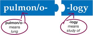
Pulmon/o-: Refers to the lung.
-logy: Means the study of.
Pulmonology: The study of the lungs and respiratory system.
Overview of the Respiratory System
Major Structures
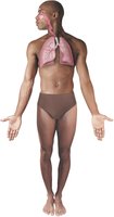
The respiratory system consists of the lungs and a series of connected structures that form the pathway for air to enter and exit the body. These structures are divided into the upper and lower respiratory tracts.
Upper respiratory tract: Nose, nasal cavity, pharynx, and larynx.
Lower respiratory tract: Trachea, bronchi, bronchioles, and lungs.
Anatomy of the Upper Respiratory System
Nose and Nasal Cavity
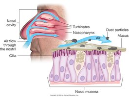
The nose is the entry point for air. The nasal cavity is divided by the septum and contains three turbinates that slow and condition the air. The nasal mucosa lines the cavity, humidifying air and trapping particles with mucus and cilia.
Turbinates: Bony structures that increase surface area and slow airflow.
Nasal mucosa: Produces mucus to trap dust and pathogens.
Cilia: Hair-like structures that move mucus and debris out of the nasal passages.
Pharynx
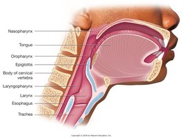
The pharynx is a muscular tube that serves as a common passageway for air and food. It is divided into three regions: nasopharynx, oropharynx, and laryngopharynx.
Nasopharynx: Uppermost part, behind the nasal cavity.
Oropharynx: Middle part, behind the oral cavity.
Laryngopharynx: Lower part, leading to the larynx and esophagus.
Anatomy of the Lower Respiratory System
Larynx (Voice Box)
The larynx remains open during breathing and contains the vocal cords, which vibrate to produce sound. During swallowing, the larynx moves upward to meet the epiglottis, preventing food from entering the airway.
Trachea (Windpipe)
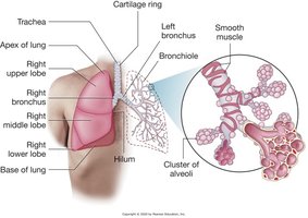
The trachea is a rigid tube supported by C-shaped cartilage rings. It conducts air to the bronchi and allows the esophagus to expand during swallowing.
Bronchi, Bronchioles, and Lungs
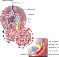
The trachea divides into right and left bronchi, which enter the lungs and branch into smaller bronchioles. At the end of bronchioles are alveoli, where gas exchange occurs.
Right lung: Three lobes (upper, middle, lower).
Left lung: Two lobes (upper, lower).
Alveoli: Microscopic air sacs for gas exchange; secrete surfactant to prevent collapse.
Thorax and Pleura
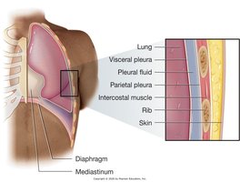
The thorax is the chest cavity, bounded by the ribs, sternum, and spine. The lungs are covered by the visceral pleura, while the parietal pleura lines the thoracic cavity. Pleural fluid lubricates the surfaces for smooth movement during breathing.
Physiology of Breathing
Mechanics of Breathing
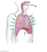
Breathing is controlled involuntarily by the brain's respiratory centers, which send signals via the phrenic nerve to the diaphragm and intercostal muscles.
Inhalation (Inspiration): Diaphragm contracts and moves downward, intercostals lift the ribs, thoracic cavity enlarges, and air flows in due to negative pressure.
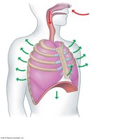
Exhalation (Expiration): Diaphragm relaxes and moves upward, intercostals lower the ribs, thoracic cavity shrinks, and air is expelled due to positive pressure.
Processes of Respiration
Respiration involves several processes:
Ventilation: Movement of air in and out of the lungs.
External respiration: Exchange of gases between alveoli and blood.
Gas transport: Movement of oxygen and carbon dioxide in the blood.
Internal respiration: Exchange of gases between blood and body cells.
Cellular respiration: Use of oxygen by cells to produce energy and carbon dioxide as a waste product.
Gas Exchange
Oxygen diffuses from alveoli into capillaries and binds to hemoglobin in red blood cells. Carbon dioxide, a metabolic waste, diffuses from blood into alveoli to be exhaled.
Common Respiratory Diseases
Upper Respiratory Infection (URI)
URIs are caused by bacteria or viruses and are highly contagious. They spread through droplets from sneezing or coughing and by contact with contaminated surfaces.

Acute Respiratory Distress Syndrome (ARDS)
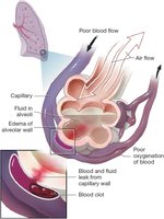
ARDS is a severe condition where alveoli fill with fluid, leading to poor oxygenation of blood. Capillary leakage and blood clots further impair gas exchange.
Atelectasis
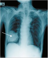
Atelectasis is the collapse of part or all of a lung, seen as a hazy area on chest x-ray. It reduces oxygen exchange and can be caused by blockage or pressure on the lung.
Cystic Fibrosis
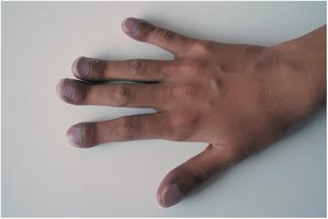
Cystic fibrosis is a genetic disorder causing thick, sticky mucus in the lungs. It leads to chronic infections, cyanosis (bluish skin), and clubbing of the fingers due to low oxygen levels.
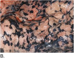
Effects of Smoking
Smoking introduces tar and other harmful substances into the lungs, leading to chronic respiratory diseases and visible deposits in lung tissue.

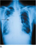
Pneumonia
Pneumonia is an infection that inflames the alveoli, causing them to fill with fluid or pus. It appears as a dense area on chest x-ray.
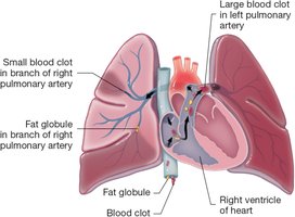
Pulmonary Embolus
A pulmonary embolus is a blood clot that travels to the lungs and blocks blood flow, causing collapse of alveoli and reduced oxygenation.
Tuberculosis
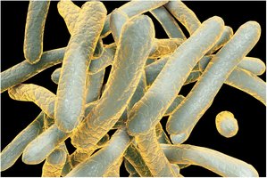
Tuberculosis is caused by Mycobacterium tuberculosis, a bacterium with a waxy coating that makes it resistant to treatment.
Diagnostic and Laboratory Procedures
Pulse Oximetry
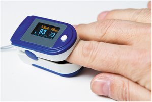
A pulse oximeter measures the percentage of hemoglobin saturated with oxygen and the pulse rate. It is a quick, noninvasive test used in many healthcare settings.
Pulmonary Function Test (PFT)
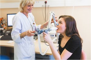
PFTs measure lung volumes and airflow to assess respiratory function, especially in chronic diseases like cystic fibrosis.
Sleep Study
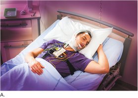
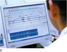
Sleep studies monitor patients overnight to diagnose sleep-related breathing disorders. Multiple sensors record physiological data for analysis.
Chest X-Ray
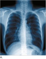
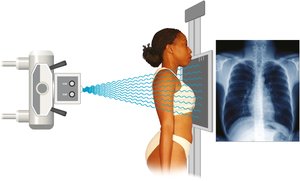
A PA (posteroanterior) chest x-ray is a standard imaging test to visualize the lungs, heart, and chest structures. It helps diagnose pneumonia, atelectasis, and other conditions.
Medical and Surgical Procedures
Auscultation and Percussion
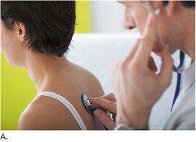
Auscultation involves listening to breath sounds with a stethoscope, while percussion involves tapping the chest to detect areas of dullness or abnormality.
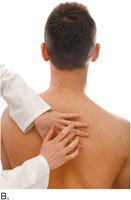
Chest Percussion Therapy
This therapy uses vibration to loosen thick mucus in the lungs, especially in cystic fibrosis patients, to aid in expectoration.
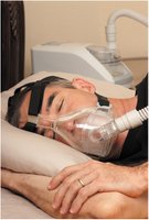
CPAP Machine
Continuous Positive Airway Pressure (CPAP) machines provide a constant flow of air to keep airways open during sleep, commonly used for sleep apnea.
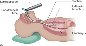
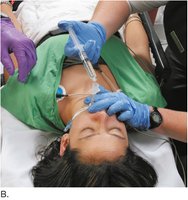
Endotracheal Intubation
An endotracheal tube is inserted into the trachea to maintain an open airway, deliver oxygen, or administer drugs in emergencies. It can be connected to a respirator for mechanical ventilation.
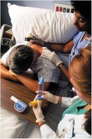
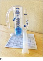
Incentive Spirometry
Incentive spirometers encourage deep breathing to expand the lungs and prevent complications after surgery or illness. Patients use the device several times daily to monitor and improve lung function.

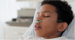
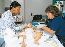
Oxygen Therapy
Oxygen can be delivered via nasal cannula or endotracheal tube to increase blood oxygen levels in patients with respiratory distress.
Drugs and Smoking Cessation
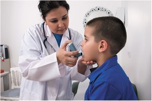
Metered-Dose Inhaler
This device delivers a premeasured dose of bronchodilator or corticosteroid medication directly to the lungs, commonly used in asthma management.
Smoking Cessation Drug
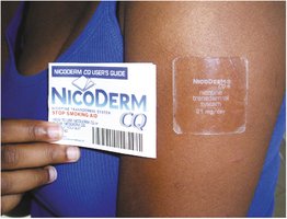
Nicotine replacement therapy, such as patches, helps patients gradually reduce dependence on nicotine and quit smoking.