 Indietro
Tissues and Organ Systems: Structure, Function, and Medical Terminology
Guida di studio - Note intelligenti
 Appunti personalizzati basati sui tuoi materiali, ampliati con definizioni chiave, esempi e contesto.
CH4: Tissues and Organ Systems
Overview of Tissues
Tissues are groups of similar cells working together to perform specific functions. There are four primary tissue classes in the human body, each with distinct roles and characteristics.
Epithelial Tissue: Maintains physical barriers, lines surfaces, and provides protection.
Connective Tissue: Connects, supports, and stores substances.
Muscle Tissue: Produces movement.
Nervous Tissue: Transmits electrical signals.
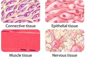
Example: The lining of the stomach is composed of epithelial tissue, while the underlying layers contain connective and muscle tissues.
Histology: Study of Microscopic Tissue Structures
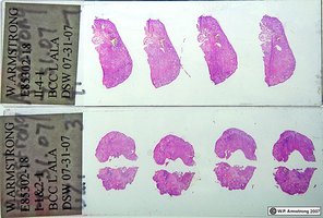
Histology is the branch of biology that studies the microscopic structure of tissues. It is essential for understanding how tissues function and how diseases affect them.
Epithelial Tissue
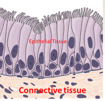
Epithelial tissue forms linings on both external and internal surfaces of the body. It is characterized by high cellularity, meaning cells are tightly packed, creating a barrier. Epithelial tissue is avascular (lacks blood vessels) and has a high regenerative capacity. Connective tissue is usually located beneath epithelial layers.
Functions: Protection, absorption, secretion, and sensation.
Locations: Skin, lining of mouth, throat, esophagus, rectum, and vagina.
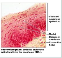
Stratified Squamous Epithelium
This type of epithelium consists of multiple layers of scaly cells, providing protection against abrasion, pathogens, and chemicals.
Glands
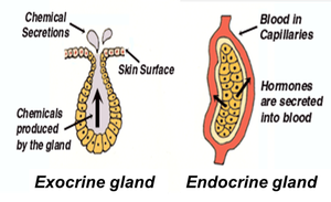
Glands are collections of epithelial cells that secrete chemical products. They are classified as:
Exocrine Glands: Secrete via ducts onto surfaces or into cavities (e.g., sweat glands).
Endocrine Glands: Ductless; secrete hormones directly into the bloodstream (e.g., thyroid gland).
Connective Tissue
Connective tissue is highly diverse, with structural composition ranging from liquid to solid. It is often found between other tissues and is usually highly vascular. Its functions include connecting, supporting, storing, and transporting substances.
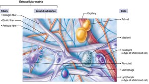
Cells: Sparse cell count; includes blast cells (immature, matrix-secreting) and cyte cells (mature, matrix-maintaining).
Ground Substance: Gelatinous material composed of proteoglycans and water.
Protein Fibers: Collagen (strength), elastic (elasticity), reticular (support netting).
Adipose Connective Tissue
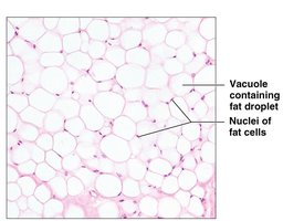
Adipose tissue contains many adipocytes (fat cells) storing triglycerides. It provides insulation and energy storage, located beneath the skin and around organs.
Dense Regular Connective Tissue
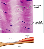
This tissue is composed of parallel collagen fibers, providing great tensile strength. It is found in tendons and ligaments.
Muscle Tissue
Muscle tissue is specialized for contraction, enabling movement. It is highly vascularized and contains many mitochondria. There are three types:
Skeletal Muscle: Long, cylindrical, multinucleate, striated cells; voluntary movement; attached to bones.
Cardiac Muscle: Branching, striated cells with intercalated discs; involuntary; found in heart wall.
Smooth Muscle: Spindle-shaped, non-striated cells; involuntary; found in hollow organs.
Nervous Tissue
Nervous tissue contains neurons (transmit electrical signals) and glial cells (support neurons). It has a poor rate of regeneration and is essential for communication and control within the body.
Organ and Organ System Level
Overview of Organ Systems
Organs are structures composed of two or more tissue types working together for specific functions. Organ systems are groups of organs that perform major body functions.
Integumentary System: Skin, nails, hair, glands; protection, sensation, thermoregulation.
Skeletal System: Bones, joints; support, protection, movement, blood cell production.
Muscular System: Skeletal, cardiac, smooth muscles; movement, heat production.
Nervous System: Brain, spinal cord, nerves; control, homeostasis.
Endocrine System: Glands; regulation of homeostasis.
Cardiovascular System: Heart, blood vessels; transport of gases, nutrients, wastes.
Lymphatic/Immune System: Lymph nodes, vessels, spleen; fluid filtration, immune response.
Respiratory System: Lungs, airways; gas exchange.
Digestive System: GI tract, liver, pancreas; digestion, absorption.
Urinary System: Kidneys, bladder; waste removal, pH, osmoregulation.
Reproductive System: Gonads, reproductive organs; offspring production.
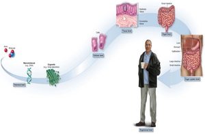
Homeostasis and Feedback Loops
Homeostasis
Homeostasis is the ability to maintain a stable internal environment despite external changes. Variables such as temperature and blood pressure oscillate around a set point.
Feedback Loops
Negative Feedback: The response negates the stimulus, returning the variable to normal. Most common type.
Positive Feedback: The response amplifies the stimulus, moving the variable further from normal. Requires external intervention to stop. Examples: childbirth, blood clotting.
Integumentary System
Layers of the Skin
The skin consists of two main layers: the epidermis (outer) and dermis (inner). The hypodermis (subcutaneous layer) is associated but not technically part of the skin.
Epidermis: Stratified squamous epithelium; produces keratin; avascular.
Dermis: Connective tissue; contains blood vessels, nerves, and sensory receptors.
Hypodermis: Adipose tissue; provides insulation and padding.
Skin Functions
Resistance to trauma and infection
Water retention
Vitamin D synthesis
Sensation
Thermoregulation
Nonverbal communication
Epidermal Sublayers
Stratum basale: Bottom layer; site of mitosis; contains melanocytes (produce melanin for pigmentation and UV protection).
Stratum corneum: Top layer; consists of dead, keratinized cells resistant to abrasion.
Dermis Components
Dermal papillae: Ridges that interlock dermis and epidermis.
Tactile corpuscles: Touch receptors.
Lamellated corpuscles: Pressure receptors.
Free nerve endings: Pain receptors.
Skin Pigments
Hemoglobin: Red pigment in blood.
Melanin: Brown pigment; protects against UV.
Collagen: White fibers; gives skin a whitish appearance.
Carotene: Orange pigment.
Bilirubin: Yellow pigment.
Skin Appendages
Sweat glands: Secrete watery solution; regulate temperature.
Sebaceous glands: Secrete sebum (oil); lubricate skin and hair.
Ceruminous glands: Produce ear wax.
Hair and nails: Protective and sensory functions.
Wound Healing
Regeneration: Minor damage; cells divide and restore tissue.
Fibrosis: Severe damage; scar tissue forms, some function lost.
Skin Cancer
ABCDE's: Asymmetry, Border irregularity, Color, Diameter (>6mm), Evolution.
Carcinogens: Substances causing mutations (e.g., UV radiation, cigarette smoke).
Benign vs. Malignant: Benign tumors do not spread; malignant tumors can metastasize.
Burn Types
1st Degree: Damages epidermis only; redness, swelling, pain.
2nd Degree: Damages epidermis and upper dermis; blisters, possible scarring.
3rd Degree: Damages entire skin thickness; risk of infection and dehydration.
Other Skin Disorders
Jaundice: Yellowing due to bilirubin.
Cyanosis: Bluish tint due to low oxygen.
Hematoma: Bruising from blood accumulation.
Blisters: Fluid-filled lesions from friction or burns.
Aging and Skin
Decrease in fluid and fibers causes sagging and wrinkles.
Increased cancer risk and slower repair rate.
Additional info: Academic context was added to clarify tissue functions, feedback mechanisms, and skin disorders.
