 Indietro
Introduction to Prokaryotic Cells: Structure, Function, and Clinical Relevance
Guida di studio - Note intelligenti
 Appunti personalizzati basati sui tuoi materiali, ampliati con definizioni chiave, esempi e contesto.
Introduction to Prokaryotic Cells
Overview of Prokaryotic Domains
Prokaryotic cells are fundamental to the study of microbiology, comprising two distinct domains: Bacteria and Archaea. These domains are separate from the domain Eukarya, which includes all eukaryotic organisms. Life on Earth is believed to have originated as prokaryotic cells approximately 3.8 billion years ago. 
General Characteristics of Prokaryotes
- Unicellular: Prokaryotes exist as single cells. - Lack a membrane-bound nucleus: Their genetic material is not enclosed within a nuclear membrane. - Lack membrane-bound organelles: Organelles such as mitochondria and endoplasmic reticulum are absent. 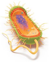
Prokaryotic Cell Morphology
Sizes of Prokaryotic Cells
Prokaryotic cells vary greatly in size, typically ranging from 0.2 to 2.0 μm in diameter. Most nutrients are obtained through diffusion, which limits cell size due to the surface area-to-volume ratio. 
Shapes and Arrangements
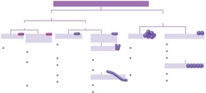
Understanding the shapes and arrangements of prokaryotes is crucial for pathogen identification. - Monomorphic: Bacteria with a single shape. - Pleomorphic: Bacteria that can take on multiple forms, aiding survival and transmission. Common shapes: - Bacilli: Rod-shaped - Cocci: Spherical - Vibrio: Comma-shaped - Stella: Star-shaped - Coccobacilli: Ovoid - Spirochetes: Spiral-shaped Common arrangements: - Diplococci: Paired cocci - Streptococci: Chains of cocci - Staphylococci: Clusters of cocci - Diplobacilli: Paired bacilli - Streptobacilli: Chains of bacilli - Palisade: Clusters of bacilli 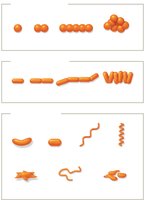
Prokaryotic Cell Division
Binary Fission
Prokaryotic cells reproduce primarily by binary fission, a process involving: 1. DNA replication 2. Cell elongation 3. Chromosome segregation to opposite ends 4. Septum formation at the midpoint 5. Separation into two daughter cells
Cell Envelope Structure
Plasma Membrane
The plasma membrane is a thin, flexible phospholipid bilayer acting as a selective barrier. It contains proteins that function as transporters, anchors, receptors, and enzymes. The membrane is also a site for metabolic reactions, including ATP synthesis. 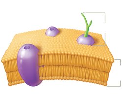
Membrane Fluidity
Membrane fluidity is essential for cell function and is influenced by: - Temperature: Higher temperatures increase fluidity; lower temperatures decrease it. - Fatty acid content: Unsaturated fatty acids enhance fluidity; saturated fatty acids make the membrane more rigid.
Archaeal vs. Bacterial Membranes
- Bacteria: Linear fatty acids, ester bonds, always bilayers. - Archaea: Long-branched fatty acids, ether bonds, can form bilayers or monolayers (especially in extreme environments). 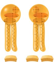
Cell Wall Structure
The cell wall provides rigidity and protection. - Bacteria: Use peptidoglycan as a core component. - Archaea: Use pseudopeptidoglycan. 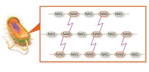
Gram Staining and Clinical Relevance
Gram-Positive vs. Gram-Negative Cell Walls
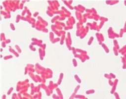
Gram-positive: - Thick peptidoglycan layer (20–80 nm) - No outer membrane - Stain purple Gram-negative: - Thin peptidoglycan layer (2–8 nm) - Outer membrane with lipopolysaccharide (LPS) - Stain red/pink 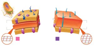
Clinical Implications
- Gram-negative bacteria are harder to kill due to their outer membrane, which acts as a selective barrier. - Gram-positive bacteria are more sensitive to agents that target peptidoglycan but retain moisture and resist mechanical stress. - Teichoic acids in Gram-positive cell walls stabilize the wall and aid in cell division.
Acid-Fast Staining
Acid-fast staining detects mycolic acid in cell walls. Acid-fast bacteria (e.g., Mycobacterium, Nocardia) appear red/pink and grow slowly due to their waxy cell wall, which also makes them resistant to drugs. 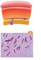
Cell Wall Variations
Mycoplasma and L-Forms
- Mycoplasma: Lack a cell wall, have a sterol-enriched plasma membrane, are pleomorphic, and often live inside other cells. - L-forms: Bacteria that lost their cell wall, contributing to persistent infections and resistance to certain stresses.
Transport Across Cell Envelope
Passive Transport
- Diffusion: Movement from high to low concentration. - Facilitated diffusion: Uses membrane proteins to move substances along their gradient. 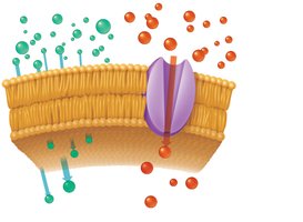
Osmosis
- Isotonic: No net water movement. - Hypertonic: Water leaves cell, causing plasmolysis. - Hypotonic: Water enters cell, causing swelling or lysis if the cell wall is damaged.
Active Transport
Prokaryotes use three main active transport mechanisms: - Primary active transport: Uses ATP to move substances against their gradient. - Secondary active transport: Uses ion gradients (symport and antiport). - Phosphotransferase systems: Group translocation, where a high-energy phosphate is transferred to the transported substance. 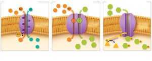
External Structures for Adhesion, Movement, and Protection
Flagella
Flagella are filamentous structures used for motility. - Gram-positive bacteria: Two rings anchor the flagellum. - Gram-negative bacteria: Four rings anchor the flagellum. 
Flagella Arrangements
- Monotrichous: Single flagellum - Lophotrichous: Tuft at one pole - Amphitrichous: Flagella at both poles - Peritrichous: Flagella all over the cell surface 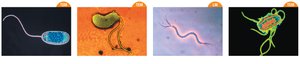
Periplasmic Flagella (Axial Filaments)
Found in spirochetes, these flagella are located between the plasma membrane and cell wall, enabling corkscrew motion. 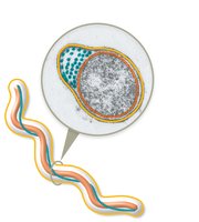
Fimbriae
Fimbriae are short, bristle-like structures that help prokaryotes adhere to surfaces and form biofilms. 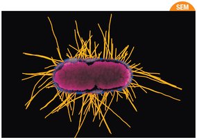
Pili
Pili are longer, less numerous than fimbriae, and aid in adhesion, movement, and gene transfer (conjugation).
Glycocalyx
The glycocalyx is a sticky, carbohydrate-rich layer that aids in adhesion, protection from desiccation, and resistance to antibiotics. - Slime layer: Unorganized, loosely associated - Capsule: Well-organized, tightly associated 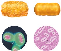
Intracellular Structures
Nucleoid
Prokaryotic DNA is organized into a single, circular chromosome located in the nucleoid region. 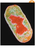
Ribosomes
Ribosomes are composed of RNA and protein, responsible for protein synthesis. Prokaryotic ribosomes are 70S, made of a 50S large subunit and a 30S small subunit. 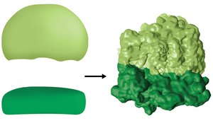
Cytoskeleton
The prokaryotic cytoskeleton consists of protein filaments that provide structural support.
Inclusion Bodies
Inclusion bodies are storage sites for substances such as carbon-fixing enzymes (carboxysomes) and magnetic iron (magnetosomes). 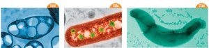
Endospores
Formation and Function
Endospores are metabolically inactive structures that allow certain bacteria to survive harsh conditions. They are highly resistant to environmental stresses and can germinate into vegetative cells when conditions improve.
Clinically Important Spore-Forming Bacteria
Genera such as Bacillus, Clostridium, and Clostridioides produce endospores, which are relevant in diseases like anthrax, tetanus, botulism, gas gangrene, and severe diarrhea.
Sporulation Process
Steps include: 1. DNA replication 2. Packaging of DNA, ribosomes, and enzymes into the spore coat 3. Formation of heat- and chemical-resistant layers 4. Release of the endospore 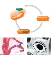
Summary Table: Gram-Positive vs. Gram-Negative Bacteria
Feature | Gram-Positive | Gram-Negative |
|---|---|---|
Peptidoglycan Layer | Thick (20–80 nm) | Thin (2–8 nm) |
Outer Membrane | Absent | Present (with LPS) |
Teichoic Acids | Present | Absent |
Staining Color | Purple | Red/Pink |
Resistance | More sensitive to peptidoglycan-targeting agents | More resistant due to outer membrane |
Additional info: This summary expands on the original notes with definitions, examples, and clinical context to provide a comprehensive, exam-ready guide for microbiology students.
