This organism causes meningitis and is transmitted mainly by the inhalation of dried, contaminated bird droppings. Infections are treated with amphotericin B and flucytosine.
Ch. 22 - Microbial Diseases of the Nervous System

Tortora14th EditionMicrobiology: An IntroductionISBN: 9780138200398Non è quello che usi tu?Cambia libro di testo
Tutti i libri di testo
Tortora 14th Edition
Ch. 22 - Microbial Diseases of the Nervous System
Problema 9
Tortora 14th Edition
Ch. 22 - Microbial Diseases of the Nervous System
Problema 9Capitolo 18, Problema 9
Use the following choices to answer the question given below:
a. Cryptococcus
b. Haemophilus
c. Listeria
d. Naegleria
e. Neisseria
Microscopic examination of cerebrospinal fluid reveals gram-positive rods.
Guida verificata passo dopo passo1
Step 1: Identify the key information given in the problem, which is that the microscopic examination of cerebrospinal fluid (CSF) reveals gram-positive rods.
Step 2: Recall the characteristics of the organisms listed in the choices, focusing on their Gram stain reaction and morphology: whether they are gram-positive or gram-negative, and whether they are rods or cocci.
Step 3: Eliminate organisms that do not match the description of gram-positive rods. For example, Cryptococcus is a yeast (not a rod), Haemophilus and Neisseria are gram-negative cocci or coccobacilli, and Naegleria is a protozoan, not a bacterium.
Step 4: Recognize that Listeria is a gram-positive rod and is known to cause meningitis, which would be consistent with its presence in cerebrospinal fluid.
Step 5: Conclude that the organism observed in the CSF as gram-positive rods is most likely Listeria, based on the process of elimination and matching characteristics.

Risposta video verificata per un problema simile:
Questa soluzione video è stata consigliata dai nostri tutor come utile per risolvere questo problema.
Durata del video:
1mConcetti chiave
Ecco i concetti essenziali che devi comprendere per rispondere correttamente alla domanda.
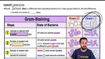
Gram Staining and Gram-Positive Rods
Gram staining differentiates bacteria by cell wall structure, with gram-positive bacteria retaining the crystal violet stain and appearing purple. Gram-positive rods are rod-shaped bacteria that stain purple, indicating a thick peptidoglycan layer. Identifying the shape and gram reaction helps narrow down possible bacterial species in infections.
Video consigliato:
Gram Stain
Common Gram-Positive Rod Pathogens in Cerebrospinal Fluid
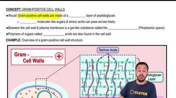
Certain gram-positive rods, like Listeria monocytogenes, are known to cause meningitis and can be found in cerebrospinal fluid. Recognizing these pathogens is crucial for diagnosis and treatment, as they differ from gram-negative or other microbial causes of CNS infections.
Video consigliato:
Gram-Positive Cell Walls
Differentiation of Microorganisms Based on Morphology and Staining
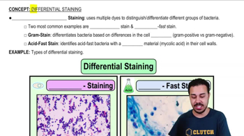
Microorganisms can be distinguished by their shape (cocci, rods), gram reaction, and other features like motility or capsule presence. For example, Neisseria is gram-negative cocci, Haemophilus is gram-negative coccobacilli, and Naegleria is a protozoan, not bacteria. This helps exclude unlikely pathogens when interpreting lab results.
Video consigliato:
Differential Staining
Pratica correlata
Domanda del libro di testo
904
views
Domanda del libro di testo
Outline the procedures for treating rabies after exposure. Outline the procedures for preventing rabies prior to exposure. What is the reason for the differences in the procedures?
829
views
Domanda del libro di testo
Provide evidence that Creutzfeldt-Jakob disease is caused by a transmissible agent.
884
views
Domanda del libro di testo
Match the following choices to the statements in questions 7 and 8:
a. antirabies antibodies
b. HDC
Used for passive immunization.
970
views
Domanda del libro di testo
Use the following choices to answer the question given below:
a. Cryptococcus
b. Haemophilus
c. Listeria
d. Naegleria
e. Neisseria
Microscopic examination of cerebrospinal fluid from a person who washes windows on a building in a large city reveals ovoid cells.
860
views