Ch. 3 - Observing Microorganisms Through a Microscope
Tortora - Microbiology: An Introduction 14th Edition
Tortora14th EditionMicrobiology: An IntroductionISBN: 9780138200398Non è quello che usi tu?Cambia libro di testo
Capitolo 3, Problema 2

Three-dimensional images of live cells can be produced with:
a. Darkfield microscopy
b. Fluorescence microscopy
c. Transmission electron microscopy
d. Confocal microscopy
e. Phase-contrast microscopy

Guida verificata passo dopo passo
1
Step 1: Understand the goal of the question, which is to identify the microscopy technique capable of producing three-dimensional images of live cells.
Step 2: Review each microscopy option and its capabilities: a) Darkfield microscopy enhances contrast but does not produce 3D images; b) Fluorescence microscopy highlights specific structures but typically produces 2D images; c) Transmission electron microscopy provides high-resolution images but requires fixed, dead samples and is 2D; d) Confocal microscopy uses laser scanning and optical sectioning to create detailed 3D images of live cells; e) Phase-contrast microscopy enhances contrast in transparent specimens but does not create 3D images.
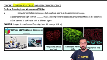
Step 3: Focus on the principle of confocal microscopy, which involves scanning a specimen with a focused laser beam and collecting light from a single focal plane, allowing the construction of 3D images by stacking multiple optical sections.
Step 4: Recognize that confocal microscopy is uniquely suited for live cell imaging in three dimensions due to its ability to optically section specimens without physical slicing.
Step 5: Conclude that the correct answer is confocal microscopy, as it is the only technique listed that can produce three-dimensional images of live cells.

Risposta video verificata per un problema simile:

Questa soluzione video è stata consigliata dai nostri tutor come utile per risolvere questo problema.
Durata del video:
2m

Concetti chiave

Ecco i concetti essenziali che devi comprendere per rispondere correttamente alla domanda.

Confocal Microscopy

Confocal microscopy uses laser light and spatial pinholes to eliminate out-of-focus light, producing sharp, high-resolution images. It allows for optical sectioning of thick specimens, enabling the reconstruction of three-dimensional images of live cells.
Video consigliato:
01:39
Confocal Scanning Laser Microscopes (CSLMs)

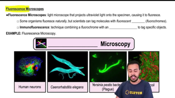
Fluorescence Microscopy

Fluorescence microscopy uses fluorescent dyes or proteins to label specific cell components, which emit light when excited by specific wavelengths. While it provides high contrast images, it typically produces two-dimensional images unless combined with techniques like confocal microscopy.
Video consigliato:
02:54
Fluorescence Microscopes

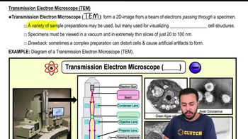
Transmission Electron Microscopy (TEM)

TEM uses electron beams transmitted through ultra-thin specimens to produce highly detailed images at the molecular level. However, it requires fixed, dead samples and cannot produce three-dimensional images of live cells.
Video consigliato:
04:23
Transmission Electron Microscope (TEM)
Pratica correlata
Domanda del libro di testo

Fill in the following blanks.

a. 1 μm = ______ m

b. 1= _______ 10⁻⁹ m

c. 1 μm = ______ nm

1563
views
Domanda del libro di testo

Assume you stain Bacillus by applying malachite green with heat and then counterstain with safranin. Through the microscope, the green structures are

a. Cell walls

b. Capsules

c. Endospores

d. Flagella

e. Impossible to identify

1899
views
Domanda del libro di testo

Looking at the cell of a photosynthetic microorganism, you observe the chloroplasts are green in brightfield microscopy and red in fluorescence microscopy. You conclude:

a. Chlorophyll is fluorescent

b. The magnification has distorted the image

c. You’re not looking at the same structure in both microscopes

d. The stain masked the green color

e. None of the above

1179
views
Domanda del libro di testo

Which type of microscope would be best to use to observe each of the following?

a. A stained bacterial smear

b. Unstained bacterial cells: the cells are small, and no detail is needed

c. Unstained live tissue when it is desirable to see some intracellular detail

d. A sample that emits light when illuminated with ultraviolet light

e. Intracellular detail of a cell that is 1μm long

f. Unstained live cells in which intracellular structures are shown in color

1799
views
Domanda del libro di testo

Carbolfuchsin can be used as a simple stain and a negative stain. As a simple stain, the pH is:

a. 2

b. Higher than the negative stain

c. Lower than the negative stain

d. The same as the negative stain

1991
views