Ch. 3 - Observing Microorganisms Through a Microscope
Tortora - Microbiology: An Introduction 14th Edition
Tortora14th EditionMicrobiology: An IntroductionISBN: 9780138200398Non è quello che usi tu?Cambia libro di testo
Capitolo 3, Problema 6

Which of the following pairs is mismatched?
a. Capsule—negative stain
b. Cell arrangement—simple stain
c. Cell size—negative stain
d. Gram stain—bacterial identification
e. None of the above

Guida verificata passo dopo passo
1
Step 1: Understand the purpose of each staining technique mentioned in the options. For example, a negative stain is used to visualize capsules and cell size by staining the background, leaving the cells or capsules clear.
Step 2: Analyze option (a): 'capsule—negative stain'. Since negative staining highlights capsules by staining the background and leaving the capsule unstained, this pair is correctly matched.
Step 3: Analyze option (b): 'cell arrangement—simple stain'. Simple stains color the cells uniformly, which helps in observing cell shape and arrangement, so this pair is also correctly matched.
Step 4: Analyze option (c): 'cell size—negative stain'. Negative staining is indeed used to observe cell size because it does not shrink or distort cells, so this pair is correctly matched as well.
Step 5: Analyze option (d): 'Gram stain—bacterial identification'. Gram staining differentiates bacteria into Gram-positive and Gram-negative groups, which is a fundamental step in bacterial identification, so this pair is correctly matched. Therefore, if all pairs are correctly matched, the answer would be (e) none of the above.

Risposta video verificata per un problema simile:

Questa soluzione video è stata consigliata dai nostri tutor come utile per risolvere questo problema.
Durata del video:
2m

Concetti chiave

Ecco i concetti essenziali che devi comprendere per rispondere correttamente alla domanda.

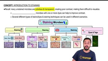
Negative Stain Technique

Negative staining uses acidic dyes that stain the background but not the cells, highlighting structures like capsules and cell size without heat-fixing. It is useful for observing delicate structures and true cell morphology.
Video consigliato:
02:10
Introduction to Staining

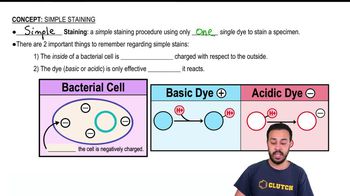
Simple Stain and Cell Arrangement

Simple staining employs a single basic dye to color cells, enhancing contrast to observe cell shape and arrangement. It helps identify how bacterial cells group together, such as chains or clusters.
Video consigliato:
03:23
Simple Staining

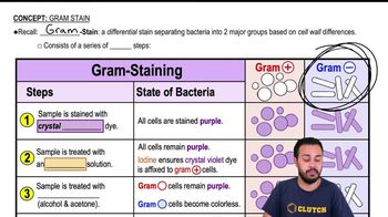
Gram Stain and Bacterial Identification

The Gram stain differentiates bacteria into Gram-positive or Gram-negative based on cell wall properties. This differential staining is fundamental for bacterial classification and guides identification and treatment.
Video consigliato:
04:57
Gram Stain
Pratica correlata
Domanda del libro di testo

What is the purpose of a counterstain in the acid-fast stain?

1768
views
Domanda del libro di testo

Why is a mordant used in the Gram stain? In the flagella stain?

2226
views
Domanda del libro di testo

The maximum magnification of a compound microscope is (a) ________; that of an electron microscope, (b) ________. The maximum resolution of a compound microscope is (c) ________; that of an electron microscope, (d) ________. One advantage of a scanning electron microscope over a transmission electron microscope is (e) ________.

1905
views
Domanda del libro di testo

Assume that you are viewing a Gram-stained field of red cocci and blue rods through the microscope. You can safely conclude that you have

a. Made a mistake in staining

b. Two different species

c. Old bacterial cells

d. Young bacterial cells

e. None of the above

1607
views
Domanda del libro di testo

Which of the following is not a functionally analogous pair of stains?

a. Nigrosin and malachite green

b. Crystal violet and carbolfuchsin

c. Safranin and methylene blue

d. Ethanol-acetone and acid-alcohol

e. All of the above pairs are functionally analogous.

1454
views
Domanda del libro di testo

Assume you stain Clostridium by applying a basic stain, carbolfuchsin, with heat, decolorizing with acid-alcohol, and counterstaining with an acidic stain, nigrosin. Through the microscope, the endospores are 1, and the cells are stained 2.

a. 1—red; 2—black

b. 1—black; 2—colorless

c. 1—colorless; 2—black

d. 1—red; 2—colorless

e. 1—black; 2—red

1649
views