 Indietro
Chapter 3: Digestion, Absorption, and Transport: The Human Body & Digestion
Guida di studio - Note intelligenti
 Appunti personalizzati basati sui tuoi materiali, ampliati con definizioni chiave, esempi e contesto.
Digestion, Absorption, and Transport
The Digestive System: Structure and Function
The digestive system is a complex network of organs responsible for breaking down food, absorbing nutrients, and eliminating waste. The gastrointestinal (GI) tract is a muscular tube approximately 20–24 feet long in adults, consisting of six main organs and several accessory organs that aid in digestion.
Organs of the GI Tract: Mouth, Pharynx, Esophagus, Stomach, Small Intestine, Large Intestine
Sphincters: Muscular valves that regulate the passage of food between organs
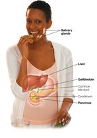
Accessory Organs: Liver, Pancreas, Gallbladder
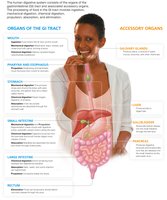
Example: The liver produces bile, which is stored in the gallbladder and released into the small intestine to aid fat digestion.
The Role of the Epiglottis and Sphincters
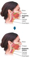
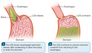
The epiglottis is a flap of tissue that prevents food from entering the trachea during swallowing. Sphincters, such as the lower esophageal sphincter (LES), control the movement of food and prevent backflow.
Epiglottis: Directs food into the esophagus, away from the airway
LES: Relaxes to allow food into the stomach, contracts to prevent reflux
Anatomy of the Stomach
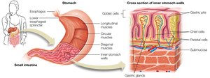
The stomach is a muscular organ that mixes and breaks down food using gastric juices. Its wall contains specialized cells that secrete enzymes and acids necessary for digestion.
Gastric cells: Chief cells (pepsinogen), parietal cells (HCl), mucous cells (mucus)
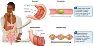
Muscle layers: Longitudinal, circular, and diagonal muscles aid in churning food
Anatomy and Function of the Small Intestine
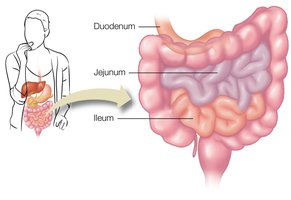
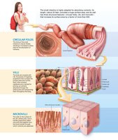
The small intestine is the primary site for nutrient absorption. Its structure maximizes surface area for efficient absorption.
Segments: Duodenum, Jejunum, Ileum
Structural features: Circular folds, villi, microvilli (increase surface area by >600 times)
The Large Intestine: Water and Nutrient Absorption
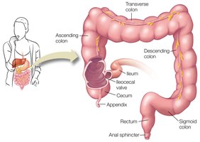
The large intestine absorbs water, sodium, potassium, and chloride, and houses the gut microbiome. It consists of the cecum, colon, and rectum.
Gut microbiome: Produces vitamins K, thiamin, riboflavin, biotin; only biotin and K are absorbed
Defecation: Final stage, under voluntary control, influenced by age, diet, and health
Accessory Organs and Digestive Juices
Accessory organs secrete substances essential for digestion.
Salivary glands: Produce saliva (water, mucus, enzymes)
Liver: Produces bile, metabolizes nutrients, detoxifies
Gallbladder: Stores and concentrates bile
Pancreas: Secretes digestive enzymes and hormones
Peristalsis and Segmentation
Peristalsis and segmentation are muscular movements that propel and mix food in the GI tract.
Peristalsis: Wave-like contractions move food forward
Segmentation: Circular muscles contract to mix and move food back and forth
Chemical Digestion: Enzymes and Conditions
Chemical digestion involves enzymes that catalyze hydrolysis reactions, breaking down macronutrients. Enzyme activity depends on substrate compatibility, pH, and temperature.
Enzyme specificity: Each enzyme acts on a specific substrate (e.g., sucrase on sucrose)
Optimal conditions: Correct pH and temperature required for activity
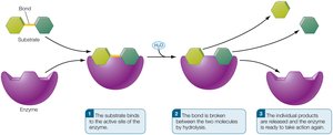
Example: Sucrase hydrolyzes sucrose into glucose and fructose.
Methods of Nutrient Absorption in the Small Intestine
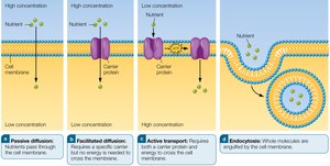
Nutrients are absorbed by four main mechanisms: passive diffusion, facilitated diffusion, active transport, and endocytosis.
Passive diffusion: Movement from high to low concentration
Facilitated diffusion: Uses carrier proteins
Active transport: Requires energy and carrier proteins
Endocytosis: Cell engulfs large molecules
Regulation of Digestion: Hormones and Nervous System
The endocrine and nervous systems coordinate digestion, absorption, and excretion. Hormones regulate secretions, motility, and hunger.
Enteric nervous system: Meshwork of nerves in GI tract, pancreas, gallbladder
Hormones: Gastrin, Secretin, CCK, GIP, Ghrelin, Peptide YY
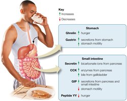
Example: CCK stimulates bile release and slows gastric motility.
Nutrient Transport: Cardiovascular and Lymphatic Systems
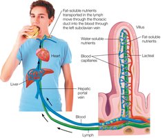
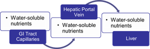
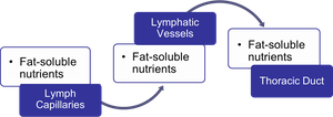
Nutrients are transported throughout the body via the cardiovascular and lymphatic systems. Water-soluble nutrients enter the bloodstream; fat-soluble nutrients enter the lymphatic system.
Water-soluble nutrients: Carbohydrates, amino acids, water-soluble vitamins (via hepatic portal vein)
Fat-soluble nutrients: Fat-soluble vitamins, long-chain fatty acids, proteins (via lymphatic vessels)
Excretory System: Waste Removal
The kidneys filter waste products from the blood, concentrating them in urine for excretion. This process is essential for maintaining homeostasis and removing metabolic byproducts.
Common Digestive Disorders
Digestive disorders range from minor to severe and can affect various parts of the GI tract.
Esophageal problems: Heartburn, esophageal cancer
Stomach disorders: Belching, gastroenteritis, ulcers (often caused by Helicobacter pylori or NSAIDs)
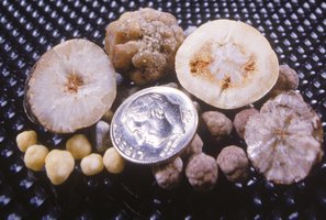
Gallbladder disease: Gallstones, risk factors include obesity and rapid weight loss
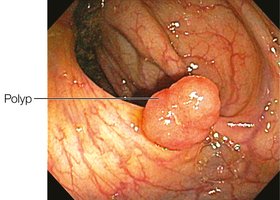
Intestinal disorders: Celiac disease, flatulence, diarrhea, constipation, hemorrhoids, IBS, IBD (ulcerative colitis, Crohn's disease), colon cancer
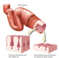
Example: Celiac disease causes flattening of intestinal villi, reducing nutrient absorption.
Fatty Liver Disease
Metabolic dysfunction–associated fatty liver disease (MAFLD) is characterized by excessive fat deposits in the liver and is associated with metabolic syndrome. It increases the risk of cardiovascular disease, cirrhosis, and GI cancers.
Risk factors: Abdominal obesity, high blood pressure, high blood glucose, abnormal blood lipids
Treatment: Weight loss, medications
