 Indietro
Heart and Neck Vessels: Structure, Function, and Clinical Assessment
Guida di studio - Note intelligenti
 Appunti personalizzati basati sui tuoi materiali, ampliati con definizioni chiave, esempi e contesto.
Heart Anatomy and Physiology
Precordium and Great Vessels
The precordium is the area of the chest overlying the heart and great vessels. Understanding its location is essential for cardiac assessment and auscultation. - Precordium: Region on the anterior chest wall where the heart is located. - Great vessels: Include the aorta, pulmonary arteries, and veins, which transport blood to and from the heart. 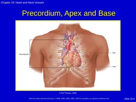
Layers of the Heart
The heart wall consists of several layers, each with distinct functions. - Pericardium: Double-walled sac that encloses the heart, providing protection and lubrication. - Epicardium: Outer layer of the heart wall. - Myocardium: Muscular middle layer responsible for contraction. - Endocardium: Inner lining of the heart chambers and valves. 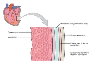
Chambers and Valves
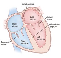
The heart contains four chambers and four main valves that regulate blood flow. - Chambers: Right atrium, right ventricle, left atrium, left ventricle. - Valves: Atrioventricular (AV) valves (tricuspid and mitral), semilunar (SL) valves (pulmonic and aortic). - Function: Valves prevent backflow and ensure unidirectional blood flow. 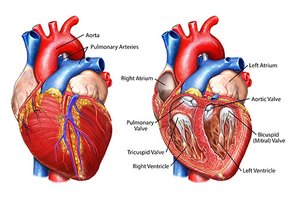
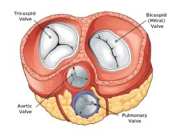
Blood Flow Through the Heart
Blood flows through the heart in a specific sequence, ensuring oxygenation and circulation. - Right side: Receives deoxygenated blood from the body and pumps it to the lungs. - Left side: Receives oxygenated blood from the lungs and pumps it to the body. 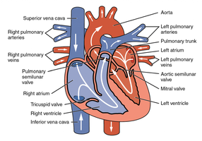
Cardiac Cycle and Heart Sounds
Cardiac Cycle
The cardiac cycle describes the rhythmic movement of blood through the heart, consisting of systole and diastole. - Diastole: Ventricles relax and fill with blood. - Systole: Heart contracts and pumps blood out.
Heart Sounds
Heart sounds are produced by the closing of valves during the cardiac cycle. - S1: First heart sound, "Lub," caused by closure of AV valves at the start of systole. - S2: Second heart sound, "Dub," caused by closure of semilunar valves at the end of systole. 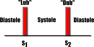
Extra Heart Sounds
- S3: Heard at the end of S2; normal in children, young adults, and pregnancy, but abnormal in older adults (may indicate heart failure). - S4: Heard just before S1; abnormal in adults, often associated with hypertension. 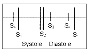
Murmurs
Murmurs are abnormal heart sounds caused by turbulent blood flow. - Characteristics: Gentle, blowing, or swooshing sound. - Grading: Ranges from Grade 1 (barely audible) to Grade 6 (loudest, stethoscope lifted off chest wall).
Pericardial Friction Rub
- Definition: High-pitched, scratchy sound caused by inflammation of the pericardium. - Best heard: At the apex and left lower sternal border with the diaphragm, patient sitting up and leaning forward.
Electrical Conduction System
Pathway of Electrical Impulses
The heart's electrical system coordinates contraction and rhythm. - SA node: Pacemaker of the heart, initiates impulse. - AV node: Delays impulse, allowing atrial contraction. - Bundle of His, bundle branches, Purkinje fibers: Conduct impulse to ventricles. 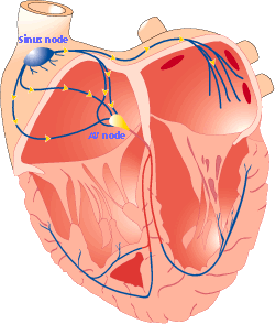
Electrocardiogram (ECG)
An ECG records the electrical activity of the heart, used to diagnose arrhythmias and other cardiac conditions. - P wave: Atrial depolarization. - QRS complex: Ventricular depolarization. - T wave: Ventricular repolarization. 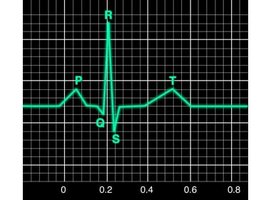
Pumping Ability and Hemodynamics
Cardiac Output and Related Terms
Cardiac output is the volume of blood pumped by the heart per minute. - Formula: $\text{Cardiac Output} = \text{Stroke Volume} \times \text{Heart Rate}$ - Stroke volume: Amount of blood ejected per beat. - Preload: Volume of blood in ventricles at end of diastole. - Afterload: Resistance the heart must overcome to eject blood. - Contractility: Strength of ventricular contraction.
Neck Vessels
Carotid Artery and Jugular Veins
The neck vessels provide important information about cardiac function. - Carotid artery: Supplies blood to the brain; assessment can reveal cardiac activity. - Jugular veins: Reflect right-sided heart activity; distention may indicate heart failure. 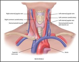
Developmental Considerations
Infants and Children
- Foramen ovale: Fetal opening between atria, closes after birth. - Ductus arteriosus: Fetal vessel connecting pulmonary artery to aorta, closes after birth. 
Pregnant Female
- Blood volume: Increases during pregnancy. - Cardiac output: Increases to meet metabolic demands. - Peripheral vasodilation: Results in decreased blood pressure.
Aging Adult
- Lifestyle impact: Smoking, alcohol, drugs, diet, exercise, stress, and genetics affect cardiac health. - Hemodynamic changes: Increased systolic BP, left ventricular wall thickness, decreased diastolic BP, dysrhythmias, ECG changes.
Clinical Assessment
Collecting Subjective Data
Subjective data includes symptoms and history relevant to cardiac function. - Chest pain or tightness: Onset, location, duration, characteristics, associated symptoms, relieving factors, medications, timing, severity. - Angina: Often described as "clenched fist" sign. 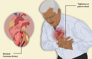
Shortness of Breath
- Dyspnea on exertion (DOE): Difficulty breathing with activity. - Paroxysmal nocturnal dyspnea (PND): Sudden nighttime breathlessness. - Orthopnea: Needing multiple pillows to breathe comfortably while lying down. 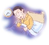

Other Symptoms
- Cough: May indicate heart failure. - Fatigue: Reduced ability to perform daily activities. - Cyanosis: Blue or ashen facial skin, sign of poor oxygenation.
Swelling and Nocturia
- Edema: Swelling of feet and legs, common in heart failure. - Nocturia: Frequent nighttime urination. 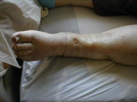
Medical and Social History
- Cardiac history: Personal and family history of heart disease. - Risk factors: Nutrition, smoking, alcohol, exercise, medications. 
Objective Data: Cardiac Exam
Carotid Arteries
- Assessment: Most accessible arteries, reflect left heart activity, assess for bruit (abnormal sound). 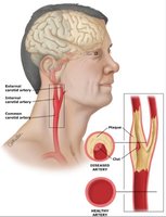
Jugular Venous Pulse
- Assessment: Reflects right heart activity, distention may indicate heart failure. 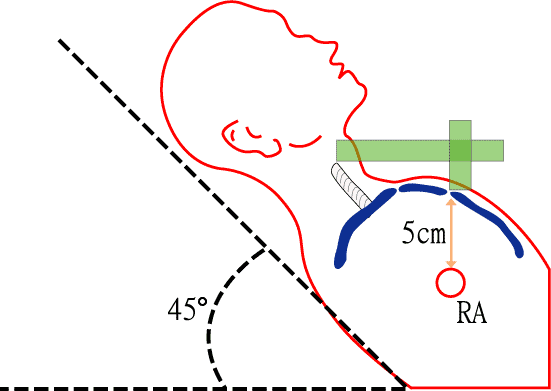
Precordium Inspection and Palpation
- Inspect: Look for pulsations, heave, or lift. - Palpate: Apical pulse and precordium.
Auscultation
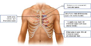
- Areas: Aortic, pulmonic, Erb’s point, tricuspid, mitral. - Technique: Begin with diaphragm, then bell; assess rate, rhythm, S1, S2, extra sounds, murmurs. 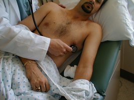
Heart Failure
Left-sided vs. Right-sided Heart Failure
Heart failure can affect either side of the heart, with distinct clinical manifestations.
Left-sided Heart Failure | Right-sided Heart Failure |
|---|---|
Dyspnea/orthopnea, cough, crackles, decreased O2 saturation, S3 heart sound, PND, tachycardia, pulmonary congestion/edema, pink frothy sputum | Dependent edema, hepatomegaly, jugular venous distention (JVD), ascites, weakness, nausea, weight gain, tachycardia |
Common Lab and Diagnostic Testing
Cardiac Biomarkers and Tests
- Lipid profile: Assesses cholesterol and triglyceride levels. - Creatinine kinase-MB (CK-MB): Indicates myocardial injury. - Troponin I: Highly specific for cardiac muscle damage. - C-reactive protein: Marker of inflammation. - B-type Natriuretic Peptide (BNP): Assesses heart failure; normal range 0-99 picograms/mL. - Electrocardiogram (ECG): Records electrical activity of the heart.
Health Promotion
Women and Heart Attack
Women may experience atypical symptoms during a heart attack. - Symptoms: Pain, breathlessness, cold sweat, anxiety, nausea, neck pain, dizziness/syncope, sleep disturbances, unusual fatigue/weakness.
Clinical Question Example
Jugular Vein Distention
Distended jugular veins in a supine patient indicate heart failure. - Correct answer: A. Heart Failure
Summary Table: Heart Sounds and Clinical Significance
Heart Sound | Timing | Clinical Significance |
|---|---|---|
S1 | Start of systole | Closure of AV valves |
S2 | End of systole | Closure of semilunar valves |
S3 | Early diastole | Normal in youth/pregnancy, abnormal in older adults (heart failure) |
S4 | Late diastole | Abnormal in adults (hypertension) |
