 Indietro
Optical Instruments: Cameras, Eyes, Magnifiers, and Microscopes
Guida di studio - Note intelligenti
 Appunti personalizzati basati sui tuoi materiali, ampliati con definizioni chiave, esempi e contesto.
Optical Instruments
Introduction
Optical instruments utilize the principles of geometric optics and lens systems to form images, magnify objects, and correct vision. This chapter explores the physics behind cameras, the human eye, magnifiers, and microscopes, focusing on how combinations of lenses create and manipulate images.
The Camera
Image Formation in Cameras
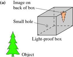
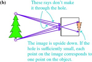
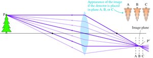
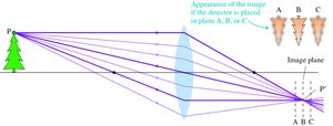
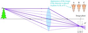
A camera projects a real image onto a plane surface, such as film or an electronic detector. The simplest camera, the pinhole camera, uses a small aperture to allow only one light ray from each point of an object to enter, forming an inverted image on the back of the box.
Pinhole Camera: Produces an inverted image; smaller holes yield sharper but dimmer images.
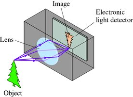
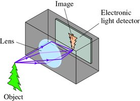
Lens Camera: Uses a converging lens to focus more light and create a sharper image.
Camera Components and Focusing
Shutter: Controls the duration of light exposure.
Diaphragm (Aperture): Adjusts the amount of light entering and affects depth of field.
Focusing: Achieved by moving the lens so the image forms sharply on the detector.
The Human Eye
Structure and Function
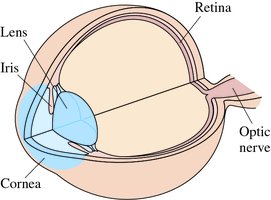
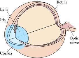
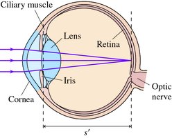
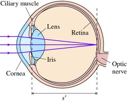
The human eye operates similarly to a camera, with an optical system (cornea and lens), a diaphragm (iris), and a light-sensitive surface (retina).
Cornea and Lens: Refract incoming light to form an image on the retina.
Iris: Regulates the amount of light entering the eye.
Retina: Detects the image, analogous to a camera's detector.
Refractive Power
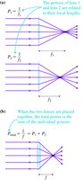
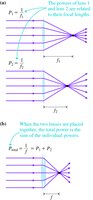
The refractive power P of a lens is the inverse of its focal length f (in meters):
Measured in diopters (D), where 1 D = 1/m. Lenses in contact have additive powers:

Focusing and Accommodation
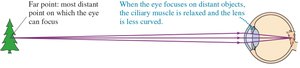
Accommodation is the process by which the eye changes the shape of its lens to focus on objects at different distances.
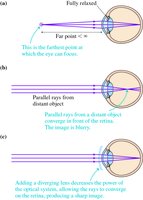
Far Point: Most distant point the relaxed eye can focus on (normally infinity).
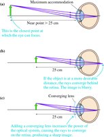
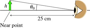
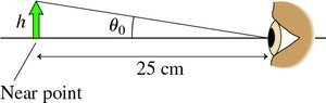
Near Point: Closest point the eye can focus on (typically 25 cm for a young adult).
Vision Defects and Correction
Presbyopia: Age-related loss of lens flexibility, reducing accommodation.
Hyperopia (Farsightedness): Near point is farther than normal; corrected with a converging lens.
Myopia (Nearsightedness): Far point is closer than normal; corrected with a diverging lens.

The Magnifier
Principle of Magnification
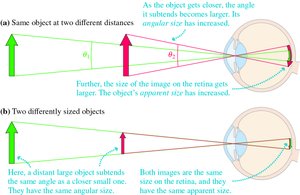
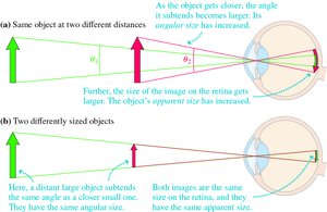
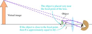
A magnifying glass allows the eye to view objects closer than the near point, increasing their angular size and apparent size on the retina.
Angular Size (θ): The angle subtended by the object at the eye; larger angular size means a larger apparent size.
Magnifying Glass: Increases the angular size by allowing the object to be placed closer to the eye.

Using a Magnifier
The maximum angular size without a lens is when the object is at the near point (25 cm):
With a magnifier (object at focal point f):
The angular magnification M is:
The Microscope
Compound Microscope Structure
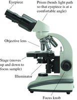
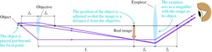
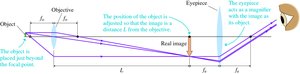
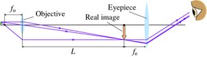
A microscope uses two lenses (objective and eyepiece) to achieve high magnification. The objective forms a real, magnified image, which is then further magnified by the eyepiece.
Objective Lens: Short focal length, creates a real image at a fixed distance (typically 160 mm).
Eyepiece: Acts as a magnifier for the real image.
Microscope Magnification
The total angular magnification M of a microscope is the product of the magnifications of the objective and eyepiece:
where L is the tube length, fo is the focal length of the objective, and fe is the focal length of the eyepiece. The negative sign indicates the image is inverted.
Summary Table: Key Optical Instrument Concepts
Instrument | Key Principle | Main Components | Magnification Equation |
|---|---|---|---|
Camera | Forms real image on detector | Lens, shutter, diaphragm, detector | |
Eye | Forms real image on retina | Cornea, lens, iris, retina | (diopters) |
Magnifier | Increases angular size | Converging lens | |
Microscope | Compound magnification | Objective, eyepiece |
Conclusion
Understanding optical instruments requires knowledge of image formation, lens combinations, and the relationship between object distance, focal length, and magnification. These principles are foundational for both physics and practical applications in technology and biology.
