 뒤로
Mass Spectrometry: Theory, Instrumentation, and Pharmaceutical Applications
스터디 가이드 - 스마트 노트
 자료에 맞춘 맞춤형 노트, 핵심 정의, 예시, 맥락을 확장해 제공합니다.
Mass Spectrometry: Principles and Applications
Introduction to Mass Spectrometry
Mass spectrometry (MS) is a powerful analytical technique used to determine the molecular weight and molecular formula of organic molecules. Unlike spectroscopic methods that rely on electromagnetic radiation, MS involves the generation, fragmentation, and separation of ions based on their mass-to-charge (m/z) ratios. When combined with other techniques such as Infrared (IR) and Nuclear Magnetic Resonance (NMR) spectroscopy, MS provides comprehensive structural information about compounds, including functional groups, carbon-hydrogen frameworks, and molecular weight.
Key Terms:
Molecular Ion (MI): The ion formed by the removal of an electron from a molecule, often denoted as M•+.
Base Peak: The most intense peak in a mass spectrum, representing the most abundant ion.
Fragmentation: The process by which molecular ions break into smaller ions and neutral fragments.
Applications: Used in pharmaceutical analysis, environmental monitoring, forensic science, and quality control.
Basic Instrumentation of Mass Spectrometry
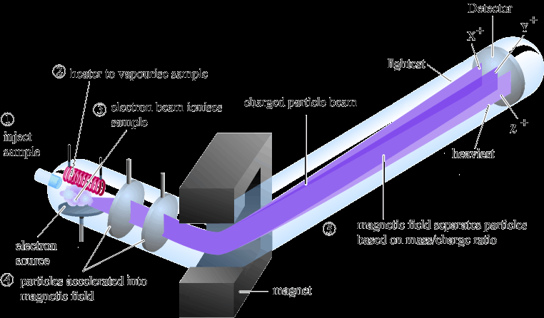
A mass spectrometer consists of several key components: a sample inlet, ionization source, mass analyzer, detector, and data system. The sample is vaporized and ionized, and the resulting ions are separated and detected based on their m/z ratios.
Sample Introduction: The sample is introduced and vaporized, often under vacuum.
Ionization: The sample is bombarded with an electron beam (typically 70 eV), generating molecular ions and fragments.
Mass Analyzer: Separates ions according to their m/z ratios using electric or magnetic fields.
Detector: Measures the abundance of separated ions, producing a mass spectrum.
Ionization Techniques
Ionization is a critical step in MS, as it determines the types of ions generated and the extent of fragmentation. Two common ionization methods are Electron Impact (EI) and Electrospray Ionization (ESI).
Electron Impact Ionization (EI):
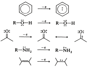
High-energy electrons (70 eV) displace an electron from the analyte, forming a radical cation (M•+).
Extensive fragmentation occurs, producing a variety of ions and neutral fragments.
Common in GC-MS and for small, volatile molecules.
Electrospray Ionization (ESI):
Soft ionization technique, often used with LC-MS.
Produces protonated (MH+) or adduct ions with minimal fragmentation.
Suitable for large, polar, and thermally labile molecules.
Common Adducts in ESI
Addition to Molecular Ion | Adduct | Comment |
|---|---|---|
M+1 | H+ | Protonation of molecule |
M+18 | NH4+ | Likely with ammonium in mobile phase |
M+22 | Na+ | Formed with traces of Na+ in mobile phase |
M+39 | K+ | Formed with traces of K+ in mobile phase |
M+41 | CH3CN+ | Formed with acetonitrile in mobile phase |
M+35 | Cl- | Formed with traces of Cl- in mobile phase |
M+45 | HCOO- | Formed with formate in mobile phase |
M+60 | CH3COO- | Formed with acetate in mobile phase |
Isotope Abundances and Mass Spectra
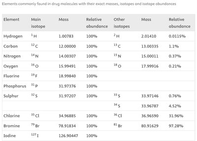
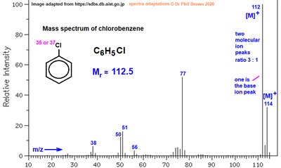
Isotopic patterns in mass spectra arise from the natural abundance of isotopes in elements such as carbon, chlorine, and bromine. These patterns are useful for identifying the presence of specific elements in a molecule.
Chlorine: 35Cl and 37Cl occur in a 3:1 ratio, producing two molecular ion peaks separated by 2 amu.
Bromine: 79Br and 81Br occur in a 1:1 ratio, also producing two peaks separated by 2 amu.
Carbon: 13C is present at about 1.1% abundance, leading to a small M+1 peak.
Fragmentation Patterns and Rules
Fragmentation in MS follows specific rules and patterns, which help in deducing the structure of the analyte. The even-electron rule states that even-electron ions tend to fragment to form other even-electron ions and neutral molecules, while odd-electron ions can fragment to form either even- or odd-electron ions.
Types of Cleavage:
Heterolytic Cleavage: Produces a cation and a neutral molecule.
Homolytic Cleavage: Produces a radical ion and a neutral radical.
Alpha (α) Cleavage: Common in compounds with heteroatoms (O, N, S), where the bond adjacent to the heteroatom is broken.
Loss of Neutral Fragments: Typical neutral losses include H2O (18 amu), CO (28 amu), CO2 (44 amu), and alkyl groups (e.g., CH3•, 15 amu).
Typical Neutral Losses in EI Fragmentation
Loss (amu) | Radicals/Neutral Fragments Lost |
|---|---|
1 | H• |
2 | H2 |
15 | CH3• |
17 | OH• or NH3 |
18 | H2O |
28 | CO |
29 | C2H5• |
31 | CH3NH2• |
43 | CH3CO• |
44 | CO2 |
45 | COOH• |
Interpretation of Mass Spectra: Examples
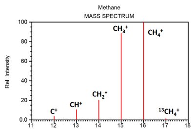
Methane (CH4)
Fragmentation sequence: CH4•+ → CH3+ → CH2+ → CH+ → C+
Parent ion peak at m/z = 16 (CH4•+), base peak at m/z = 15 (CH3+).
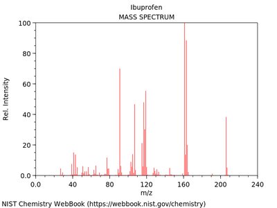
Ibuprofen
Molecular ion at m/z = 206.
Major fragments: m/z = 163 (loss of propyl group), m/z = 161 (base peak, loss of COOH), m/z = 119 (loss of CO2 from 163).
Tropylium ion formation is observed.
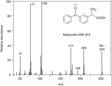
Ketoprofen
Molecular ion at m/z = 254.
Major fragments: m/z = 209, 177, 105, 77.
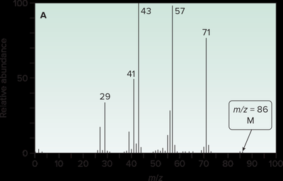
Alkyl Chains: 2-Methylpentane vs. 2,2-Dimethylbutane
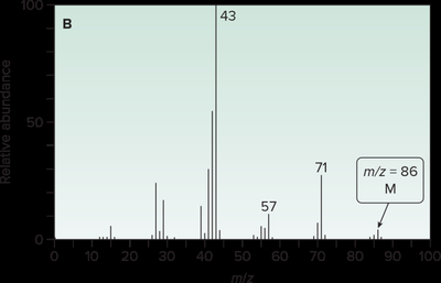
2-Methylpentane: Base peak at m/z = 43 (2° carbocation).
2,2-Dimethylbutane: Base peak at m/z = 57 (3° carbocation).
Fragmentation of Functional Groups
Aldehydes and Ketones: Undergo α-cleavage next to the carbonyl group, forming resonance-stabilized acylium cations.
Alcohols: Fragment via α-cleavage or dehydration, forming carbocations or alkenic cationic radicals.
Amines: Undergo α-cleavage between the alkyl group and the nitrogen, forming resonance-stabilized carbocations.
Esters and Amides: May undergo McLafferty rearrangement if a γ-hydrogen is present, resulting in the formation of a neutral alkene and a radical ion with m/z = +1.
Coupled Chromatography-Mass Spectrometry Systems
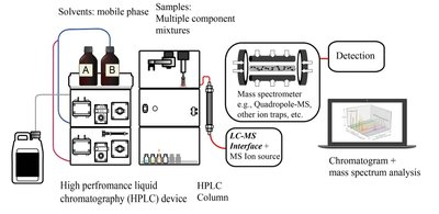
To analyze mixtures, MS is often coupled with chromatographic techniques such as Gas Chromatography (GC-MS) or Liquid Chromatography (LC-MS). Chromatography separates the mixture into individual components, which are then analyzed by MS.
GC-MS: Used for volatile compounds, environmental pollutants, food analysis, forensic toxicology, and pharmaceutical impurity identification.
LC-MS: Used for non-volatile, polar, or thermally labile compounds, biomedical analysis, environmental monitoring, and pharmaceutical quality control.
Case Studies: Interpretation of Mass Spectra
4-Chlorobenzamide (C7H6ClNO)
Major peaks: m/z = 139, 141, 157, 155, 111, 113, 44.
Peaks at m/z 139 and 141 correspond to the molecular ion peaks due to the presence of 35Cl and 37Cl isotopes (3:1 ratio).
Methyl Salicylate
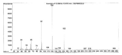
Base peak and parent ion peak can be identified from the spectrum.
Fragmentation to m/z = 120 and 92 explained by loss of specific groups (e.g., methoxy or carboxyl).
3-Methoxy Benzoic Acid
Base peak identification and explanation of peaks at m/z = 135 and 107 based on fragmentation pathways.
Summary
Mass spectrometry is essential for molecular weight determination, structural elucidation, and impurity analysis in pharmaceuticals.
Understanding ionization methods, fragmentation patterns, and isotopic abundances is crucial for interpreting mass spectra.
Coupling MS with chromatographic techniques enhances its application to complex mixtures in various scientific fields.
