 뒤로
LEC 1 SLIDES
스터디 가이드 - 스마트 노트
 자료에 맞춘 맞춤형 노트, 핵심 정의, 예시, 맥락을 확장해 제공합니다.
Anatomical Terminology
Introduction to Anatomical Language
Anatomy uses a specialized language to describe the structures, locations, and relationships within the human body. Mastery of this terminology is essential for clear communication and understanding in the field of anatomy and physiology.
Microscopic Anatomy: Includes cytology (study of cells) and histology (study of tissues).
Gross Anatomy: Study of structures visible to the naked eye.
Anatomical Position: The standard reference position for anatomical descriptions.

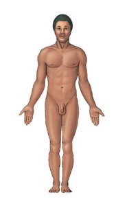
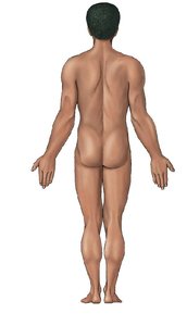
Directional Terms
Directional terms are used to describe the locations of structures relative to other structures or locations in the body.
Cranial (Cephalic): Toward the head
Caudal: Toward the tail
Anterior (Ventral): The front, before
Posterior (Dorsal): The back, behind
Proximal: Toward a base
Distal: Away from the base
Lateral: Away from midline
Medial: Toward the midline
Superficial: At or close to body surface
Deep: Further from surface, toward interior
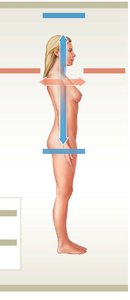
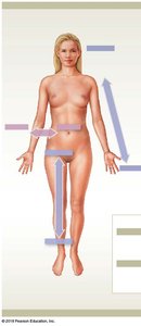
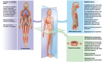
Anatomical Planes
The body can be divided into sections using three principal planes: sagittal, frontal (coronal), and transverse (horizontal).
Sagittal Plane: Divides the body into left and right portions.
Frontal (Coronal) Plane: Divides the body into anterior and posterior portions.
Transverse (Horizontal) Plane: Divides the body into superior and inferior portions.
Body Cavities
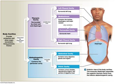
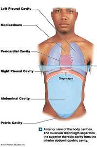
Body cavities provide protection to internal organs and allow for changes in organ shape and size.
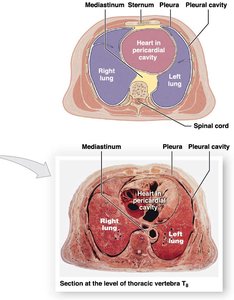
Thoracic Cavity: Contains pleural cavities (lungs), pericardial cavity (heart), and mediastinum.
Abdominopelvic Cavity: Contains abdominal cavity (digestive organs) and pelvic cavity (urinary bladder, reproductive organs).
Regions and Landmarks
Specific regions and landmarks are used to describe locations on the body with precision.
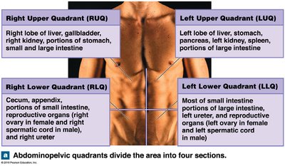
Quadrants: The abdominopelvic area is divided into four quadrants (RUQ, LUQ, RLQ, LLQ).
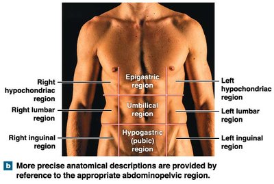
Regions: More precise descriptions use nine regions (e.g., epigastric, umbilical, hypogastric).
Tissues and Early Embryology
Early Embryology and Formation of Tissues
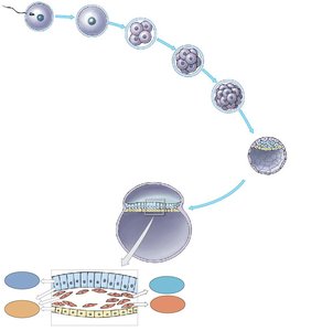
Embryology describes the development of tissues from a single fertilized cell through successive stages.
Fertilization: Sperm and egg unite to form a zygote.
Cleavage: Rapid cell divisions create a blastocyst.
Blastocyst: Contains trophoblast (forms placenta) and inner cell mass (forms embryo).
Germ Layers: Endoderm, mesoderm, and ectoderm give rise to all tissues and organs.
Primary Tissue Types
All cells in the body are classified into four primary tissue types, each with distinct functions and characteristics.
Epithelial Tissue: Covers surfaces, lines cavities, forms glands.
Connective Tissue: Provides support, protection, and energy storage.
Muscular Tissue: Specialized for contraction and movement.
Neural Tissue: Specialized for rapid communication via electrical signals.
Epithelial Tissue
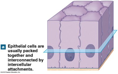
Epithelial tissue is characterized by closely packed cells with distinct polarity and specialized attachments.
Cellularity: Cells are tightly bound with minimal intercellular space.
Polarity: Apical (exposed) and basal (attached) surfaces.
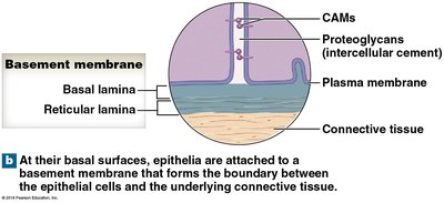
Attachment: Basal surface attached to basal lamina; cells connected by various junctions.
Types of Epithelium
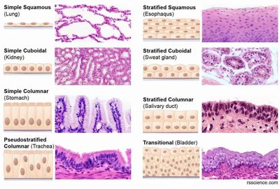
Epithelium is classified by cell shape and number of layers.
Simple: One layer of cells (squamous, cuboidal, columnar, pseudostratified columnar).
Stratified: Multiple layers (squamous, cuboidal, columnar, transitional).
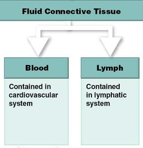
Connective Tissue
Connective tissue is defined by its cells, fibers, and matrix, and is the most histologically variable tissue type.
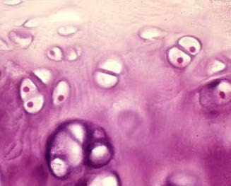
Specialized Cells: Vary by tissue type (e.g., fibroblasts, chondrocytes).
Extracellular Protein Fibers: Collagen, elastin, reticular fibers.
Matrix: Combination of fibers and ground substance (e.g., plasma in blood).
Functions: Structural support, protection, energy storage.
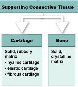
The Integumentary System
Structure and Function of the Skin
The integumentary system consists of the skin and its accessory structures, providing protection, sensation, and regulation.
Epidermis: Outermost layer, composed of epithelial cells.
Dermis: Middle layer, contains connective tissue, blood vessels, nerves.
Hypodermis (Subcutaneous Layer): Deepest layer, contains adipose tissue and binds skin to underlying structures.
Skin Cancers
Skin cancers arise from different cell types within the epidermis and vary in severity and prognosis.
Basal Cell Carcinoma: Most common, slow-growing, arises from basal cells.
Squamous Cell Carcinoma: Less common, arises from squamous cells, generally slow-growing.
Melanoma: Rare but most serious, arises from melanocytes, highly metastatic.
Dermis: Papillary and Reticular Layers
The dermis is divided into two layers, each with distinct connective tissue types and functions.
Papillary Layer: Loose areolar connective tissue, contains fibroblasts, blood vessels, nerve cells.
Reticular Layer: Dense irregular connective tissue, provides strength and elasticity.
Subcutaneous Layer (Hypodermis)
The hypodermis is the deepest layer of skin, containing adipose tissue, blood vessels, and nerves.
Functions: Insulation, energy storage, binding skin to underlying tissues.
Accessory Structures: Hair and Sebaceous Glands
Hair and sebaceous glands are accessory structures of the skin, each with specialized functions.
Hair Follicle: Produces hair via keratinization; contains papilla and bulb.
Sebaceous Gland: Produces oily sebum, released via holocrine secretion.
Accessory Structures: Nails
Nails are protective structures formed from keratinized epithelial cells.
Skin Repair and Regeneration
Skin repair involves epithelial wound healing and, in some cases, regeneration of deeper structures such as fingertips.
Factors Affecting Regeneration: Age, treatment, and amputation plane.
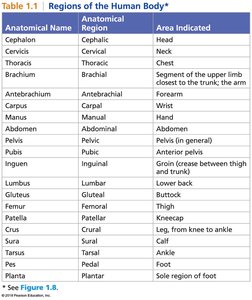
Summary Table: Regions of the Human Body
Anatomical Name | Anatomical Region | Area Indicated |
|---|---|---|
Cephalon | Cephalic | Head |
Cervicis | Cervical | Neck |
Thoracis | Thoracic | Chest |
Brachium | Brachial | Upper arm |
Antebrachium | Antebrachial | Forearm |
Carpus | Carpal | Wrist |
Manus | Manual | Hand |
Abdomen | Abdominal | Abdomen |
Pelvis | Pelvic | Pelvis |
Pubis | Pubic | Anterior pelvis |
Inguen | Inguinal | Groin |
Lumbus | Lumbar | Lower back |
Gluteus | Gluteal | Buttock |
Femur | Femoral | Thigh |
Patella | Patellar | Kneecap |
Crus | Crural | Leg |
Sura | Sural | Calf |
Pedes | Pedal | Foot |
Planta | Plantar | Sole of foot |
Summary Table: Types of Connective Tissue
Type | Subtypes | Main Features |
|---|---|---|
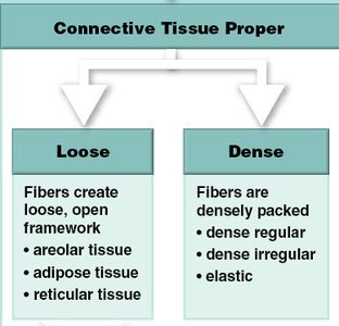
Connective Tissue Proper | Loose (areolar, adipose, reticular), Dense (regular, irregular, elastic) | Fibers and cells in variable arrangements |
Fluid Connective Tissue | Blood, Lymph | Liquid matrix |
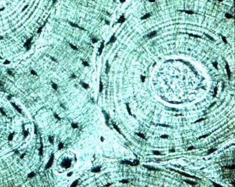
Supporting Connective Tissue | Cartilage (hyaline, elastic, fibrous), Bone | Solid matrix |
Summary Table: Types of Epithelium
Type | Location | Function |
|---|---|---|
Simple Squamous | Lung | Diffusion, filtration |
Simple Cuboidal | Kidney | Secretion, absorption |
Simple Columnar | Stomach | Absorption, secretion |
Pseudostratified Columnar | Trachea | Secretion, movement of mucus |
Stratified Squamous | Esophagus | Protection |
Stratified Cuboidal | Sweat gland | Protection |
Stratified Columnar | Salivary duct | Protection, secretion |
Transitional | Bladder | Stretching |
Additional info: Academic context was added to clarify tissue types, embryology, and anatomical terminology for completeness and exam preparation.
