 뒤로
Anatomy & Physiology Study Guide: Bones of the Human Skeleton
스터디 가이드 - 스마트 노트
 자료에 맞춘 맞춤형 노트, 핵심 정의, 예시, 맥락을 확장해 제공합니다.
Bones of the Human Skeleton
Mandible and Associated Structures
The mandible is the lower jawbone and is the largest, strongest bone of the face. It plays a crucial role in mastication (chewing) and forms the lower jawline.
Alveolar Process: The ridge containing the sockets for the lower teeth.
Mandibular Condyle: Articulates with the temporal bone to form the temporomandibular joint.
Mandibular Foramen: Passage for nerves and blood vessels to the lower teeth.
Mental Foramen: Opening for nerves and vessels to the chin and lower lip.
Ramus: Vertical extension of the mandible.
Body: Horizontal portion holding the teeth.
Scapula (Shoulder Blade)
The scapula is a flat, triangular bone located on the posterior thoracic wall. It connects the humerus (upper arm bone) with the clavicle (collarbone).
Acromion: Extension forming the highest point of the shoulder.
Coracoid Process: Provides attachment for muscles.
Spine: Prominent ridge on the posterior surface.
Glenoid Cavity: Articulates with the head of the humerus.
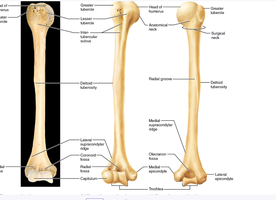
Humerus (Upper Arm Bone)
The humerus is the bone of the upper arm, connecting the shoulder to the elbow.
Head: Articulates with the glenoid cavity of the scapula.
Neck: Anatomical and surgical necks are important landmarks.
Greater and Lesser Tubercle: Sites for muscle attachment.
Deltoid Tuberosity: Attachment for the deltoid muscle.
Capitulum and Trochlea: Articulate with the radius and ulna at the elbow.
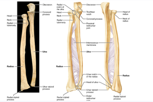
Radius and Ulna (Forearm Bones)
The radius and ulna are the two bones of the forearm. The radius is lateral (thumb side), and the ulna is medial (little finger side).
Olecranon: The point of the elbow, part of the ulna.
Coronoid Process: Anterior projection of the ulna.
Radial Tuberosity: Attachment for the biceps muscle.
Styloid Processes: Distal projections for ligament attachment.
Interosseous Membrane: Connects the radius and ulna.
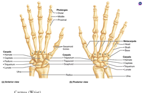
Hand Bones: Carpals, Metacarpals, and Phalanges
The hand consists of three groups of bones: carpals (wrist), metacarpals (palm), and phalanges (fingers).
Carpals: Eight small bones forming the wrist.
Metacarpals: Five bones forming the palm, numbered I-V from thumb to little finger.
Phalanges: Fourteen bones forming the fingers (proximal, middle, distal).
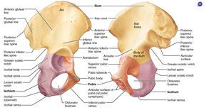
Os Coxae (Pelvic Bone)
The os coxae forms the pelvis and consists of three fused bones: ilium, ischium, and pubis. It supports the weight of the upper body and protects pelvic organs.
Iliac Crest: Superior border of the ilium.
Acetabulum: Socket for the head of the femur.
Obturator Foramen: Large opening for nerves and blood vessels.
Ischial Tuberosity: Supports weight when sitting.
Anterior/Posterior Superior/Inferior Iliac Spine: Muscle attachment points.
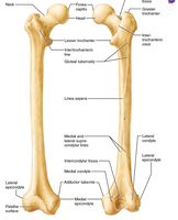
Femur (Thigh Bone)
The femur is the longest and strongest bone in the body, forming the thigh and articulating with the pelvis and tibia.
Head: Fits into the acetabulum of the pelvis.
Neck: Connects the head to the shaft.
Greater and Lesser Trochanter: Muscle attachment sites.
Medial and Lateral Condyles: Articulate with the tibia.
Linea Aspera: Ridge for muscle attachment.
Tibia and Fibula (Lower Leg Bones)
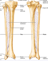
The tibia and fibula are the bones of the lower leg. The tibia is larger and medial, bearing most of the weight, while the fibula is lateral and slender.
Medial and Lateral Condyles: Articulate with the femur.
Tibial Tuberosity: Attachment for the patellar ligament.
Medial Malleolus: Distal projection of the tibia.
Lateral Malleolus: Distal projection of the fibula.
Interosseous Membrane: Connects tibia and fibula.
Foot Bones: Tarsals, Metatarsals, and Phalanges
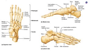
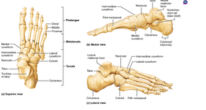
The foot is composed of tarsals (ankle), metatarsals (foot), and phalanges (toes). These bones provide support and mobility.
Tarsals: Seven bones forming the ankle, including the talus and calcaneus.
Metatarsals: Five bones forming the foot, numbered I-V.
Phalanges: Fourteen bones forming the toes.
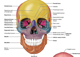
Skull: Facial and Cranial Bones
The skull consists of cranial and facial bones that protect the brain and support facial structures.
Parietal, Frontal, Temporal, Occipital Bones: Form the cranium.
Maxilla, Mandible, Zygomatic, Nasal Bones: Form the face.
Foramina: Openings for nerves and blood vessels.
Summary Table: Major Bones and Their Features
Bone | Key Features | Function |
|---|---|---|
Mandible | Alveolar process, condyle, foramen | Chewing, jaw movement |
Scapula | Acromion, coracoid, spine, glenoid cavity | Shoulder articulation |
Humerus | Head, tubercles, condyles | Arm movement |
Radius/Ulna | Styloid process, olecranon, interosseous membrane | Forearm rotation |
Os Coxae | Iliac crest, acetabulum, obturator foramen | Pelvic support |
Femur | Head, neck, trochanters, condyles | Thigh movement |
Tibia/Fibula | Condyles, malleolus, tuberosity | Leg support |
Hand/Foot | Carpals/tarsals, metacarpals/metatarsals, phalanges | Manipulation, locomotion |
Skull | Cranial/facial bones, foramina | Protection, support |
Additional info:
All bones are classified as either axial (skull, vertebral column, rib cage) or appendicular (limbs, girdles).
Joints are formed where two bones meet, allowing for movement and flexibility.
Bone markings (processes, foramina, tuberosities) are important for muscle attachment and passage of nerves/vessels.
