 뒤로
뒤로Anatomy & Physiology Study Guide: Chapter 4 – Tissues
스터디 가이드 - 스마트 노트
 자료에 맞춘 맞춤형 노트, 핵심 정의, 예시, 맥락을 확장해 제공합니다.
Foundations of Histology
Definition and Levels of Organization
Histology is the scientific study of tissues, which are groups of cells related in structure and function, plus their extracellular matrix (ECM), working together to perform common functions. Tissues are the third of six levels of structural organization in the human body: Chemical → Cellular → Tissue → Organ → Organ System → Organism.
Chemical Level: Atoms and molecules form the basis of all living structures.
Cellular Level: Molecules combine to form cells, the smallest living units.
Tissue Level: Similar cells and their ECM work together.
Organ Level: Two or more tissue types combine to form organs.
Organ System Level: Organs work together in systems.
Organism Level: The complete living individual.
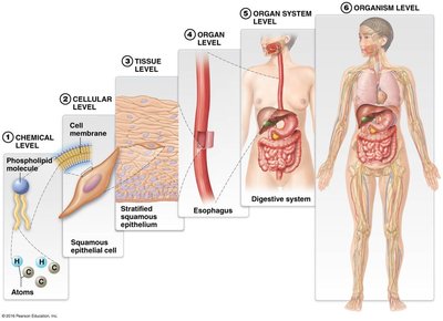
Example: The digestive system illustrates all levels, from atoms to the whole organism.
Tissues in Organs: The Trachea Example
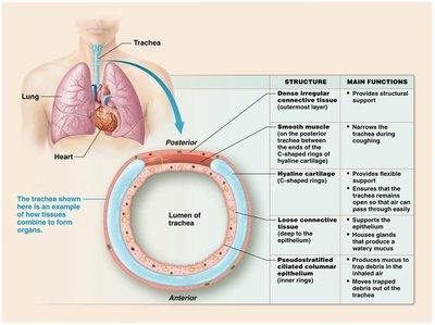
Organs are composed of multiple tissue types, each contributing to the organ's function. The trachea demonstrates how four tissue types combine:
Pseudostratified ciliated columnar epithelium: Produces mucus and moves debris out of the airway.
Loose connective tissue: Supports epithelium and houses glands.
Hyaline cartilage: Provides flexible support and keeps the airway open.
Smooth muscle: Narrows the trachea during coughing.
Dense irregular connective tissue: Provides structural support.
Major Tissue Types
Comparison of the Four Major Tissue Types
The human body contains four major tissue types, each with distinct features and functions:
Epithelial Tissue: Covers and lines surfaces; forms glands; tightly packed cells with little ECM.
Connective Tissue: Binds, supports, protects, and transports; cells scattered in abundant ECM.
Muscle Tissue: Contracts to generate force; cells packed closely with little ECM.
Nervous Tissue: Generates and transmits messages; neurons and neuroglial cells in unique ECM.
Key distinguishing feature: The amount and composition of ECM varies greatly among tissue types.
Extracellular Matrix (ECM)
Components and Functions
The ECM is the material outside cells, consisting of ground substance and protein fibers. It provides structural support, binds cells, and influences cell behavior.
Ground Substance: Water, ions, glycosaminoglycans (GAGs), proteoglycans, adhesive glycoproteins.
Protein Fibers: Collagen (strength), elastic (stretch/recoil), reticular (supportive mesh).
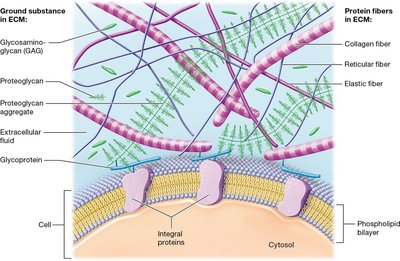
Example: Collagen fibers resist stretching, while elastic fibers allow tissues to stretch and recoil.
Cell Junctions
Types of Junctions
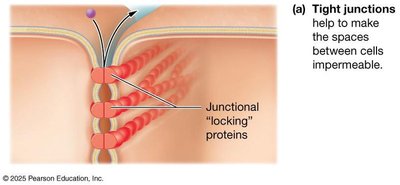
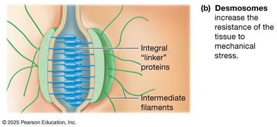
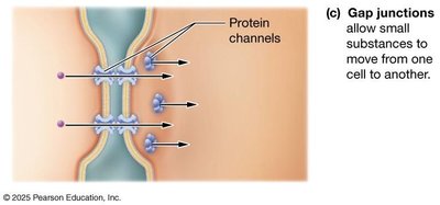
Cells in tissues are united by cell-adhesion molecules (CAMs) and cell junctions. The three main junctions are:
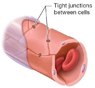
Tight Junctions: Seal spaces between cells, making them impermeable.
Desmosomes: Provide mechanical strength by linking cells.
Gap Junctions: Allow communication by forming channels for small substances.
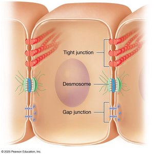
Example: Tight junctions in blood vessels prevent substances from leaking between cells.
Epithelial Tissue
Classification and Identification
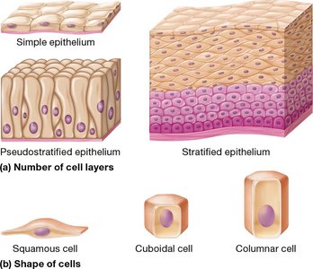
Epithelial tissues are classified by the number of cell layers (simple, stratified, pseudostratified) and cell shape (squamous, cuboidal, columnar).
Simple: One layer; good for diffusion and absorption.
Stratified: Multiple layers; good for protection.
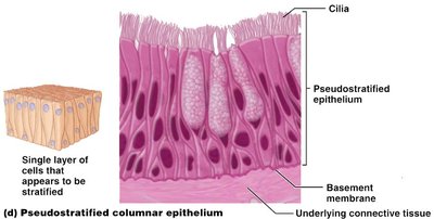
Pseudostratified: Appears stratified but is a single layer.
Types of Epithelial Tissue
Each epithelial type has a unique structure, function, and location:
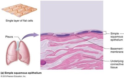
Simple squamous: Thin, flat cells; gas/fluid exchange; found in lungs, blood vessels.
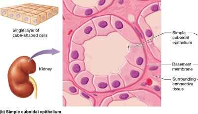
Simple cuboidal: Cube-shaped cells; absorption/secretion; found in kidney tubules, glands.
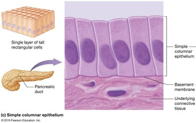
Simple columnar: Tall cells; absorption/secretion; found in digestive tract, uterine tube.
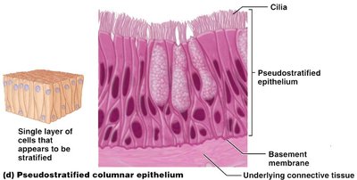
Pseudostratified ciliated columnar: Single layer with cilia/goblet cells; mucus secretion; found in respiratory tract.
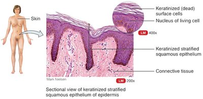
Keratinized stratified squamous: Multiple layers, dead keratin-filled cells; protection; found in skin.
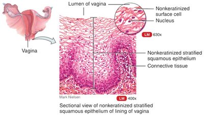
Nonkeratinized stratified squamous: Multiple layers, living cells; protection; found in mouth, vagina.
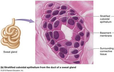
Stratified cuboidal: Two or more layers of cubes; absorption/secretion; found in sweat glands.
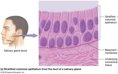
Stratified columnar: Two or more layers, columnar top; protection; found in salivary gland ducts.
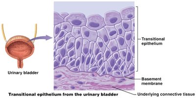
Transitional: Multiple layers, dome-shaped top; stretches; found in urinary bladder.
Glands
Exocrine vs. Endocrine Glands
Glands are specialized epithelial structures that secrete substances. They are classified as:
Endocrine: Ductless; secrete hormones into interstitial fluid and blood; act on distant targets.
Exocrine: Usually have ducts; secrete onto epithelial surfaces; act locally.
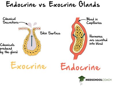
Example: Sweat glands are exocrine; thyroid gland is endocrine.
Connective Tissue
Types and Functions
Connective tissues are diverse, with functions determined by their ECM:
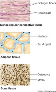
Dense regular connective tissue: Parallel collagen fibers; strength in one direction.
Adipose tissue: Fat storage; insulation and cushioning.
Bone tissue: Hard matrix; support and protection.
Muscle Tissue
Types and Characteristics
Muscle tissue is specialized for contraction and movement. The three types are:
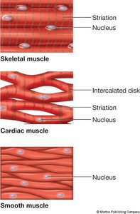
Skeletal muscle: Long, striated, multinucleated; voluntary movement.
Cardiac muscle: Short, branching, striated, one nucleus, intercalated discs; involuntary, heart.
Smooth muscle: Spindle-shaped, no striations, one nucleus; involuntary, walls of hollow organs.
Nervous Tissue
Neurons and Neuroglial Cells
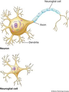
Nervous tissue consists of neurons (excitable cells that transmit electrical signals) and neuroglial cells (support, protect, and nourish neurons).
Neuron: Cell body, axon, dendrites; cannot usually divide.
Neuroglial cell: Smaller, can divide; various supportive functions.
Summary Table: Epithelial Tissue Types
Epithelium | Key Structure | Function | Location |
|---|---|---|---|
Simple squamous | 1 layer flat cells | Gas/fluid exchange | Lung air sacs, blood vessels |
Simple cuboidal | 1 layer cube cells | Absorption, secretion | Kidney tubules, glands |
Simple columnar | 1 layer tall cells | Absorption, secretion | Digestive tract, uterine tube |
Pseudostratified ciliated columnar | 1 layer, cilia/goblet cells | Mucus secretion | Respiratory tract |
Keratinized stratified squamous | Many layers, dead keratin cells | Protection | Skin |
Nonkeratinized stratified squamous | Many layers, living cells | Protection | Mouth, vagina |
Stratified cuboidal | 2+ layers cube cells | Absorption, secretion | Sweat glands |
Stratified columnar | 2+ layers, columnar top | Protection | Salivary gland ducts |
Transitional | Many layers, dome top | Stretching | Urinary bladder |
Summary Table: Connective Tissue Types
Type | ECM | Cells | Function | Location |
|---|---|---|---|---|
Dense regular | Parallel collagen | Fibroblasts | Strength | Tendons, ligaments |
Adipose | Sparse, fat droplets | Adipocytes | Fat storage | Hypodermis, organs |
Bone | Hard matrix | Osteocytes | Support, protection | Bones |
Summary Table: Muscle Tissue Types
Type | Cell Shape | Striations | Nuclei | Control | Location |
|---|---|---|---|---|---|
Skeletal | Long, cylindrical | Yes | Many | Voluntary | Skeleton |
Cardiac | Short, branching | Yes | One | Involuntary | Heart |
Smooth | Spindle-shaped | No | One | Involuntary | Hollow organs |
Summary Table: Nervous Tissue
Cell Type | Main Job | Structure | Division Ability |
|---|---|---|---|
Neuron | Transmit signals | Cell body, axon, dendrites | No |
Neuroglial cell | Support neurons | Small, various types | Yes |
Additional info: These notes expand on the original study guide by providing full academic explanations, summary tables, and relevant images for each tissue type, making the content self-contained and suitable for exam preparation.
