 뒤로
neuro 7
스터디 가이드 - 스마트 노트
 자료에 맞춘 맞춤형 노트, 핵심 정의, 예시, 맥락을 확장해 제공합니다.
Anatomy of the Central Nervous System
Gray Matter vs. White Matter
The central nervous system (CNS) is composed of the brain and spinal cord, which are structurally divided into gray matter and white matter. Understanding these divisions is fundamental to grasping CNS function.
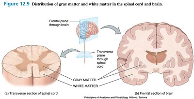
Gray Matter: Consists of unmyelinated somas, dendrites, and axons. It is the site of synaptic integration and processing.
White Matter: Composed mainly of myelinated axons, responsible for transmitting signals between different CNS regions.
Distribution: In the spinal cord, gray matter is central, surrounded by white matter; in the brain, gray matter forms the cortex and some internal nuclei, while white matter lies beneath.
Spinal Cord Structure and Function
The spinal cord is the major pathway for information flow between the brain and the peripheral body. It is divided into regions and segments, each giving rise to spinal nerves.
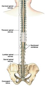
Regions: Cervical (8), Thoracic (12), Lumbar (5), Sacral (5), Coccygeal (1).
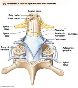
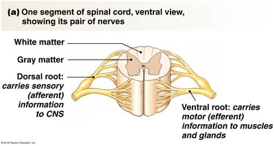
Spinal Nerves: Each segment produces a bilateral pair of nerves, which split into dorsal (sensory) and ventral (motor) roots.
Nuclei vs. Ganglia: Nuclei are clusters of cell bodies in the CNS; ganglia are clusters in the PNS.
Spinal Cord Segments and Roots
Dorsal Root: Carries sensory (afferent) information to the CNS.
Ventral Root: Carries motor (efferent) information to muscles and glands.
Gray Matter in the Spinal Cord
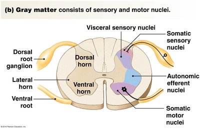
Gray matter in the spinal cord consists of sensory and motor nuclei, organized into horns.
Dorsal Horn: Contains sensory nuclei (somatic and visceral).
Ventral Horn: Contains motor nuclei (somatic and autonomic).
Lateral Horn: Present in thoracic and upper lumbar regions, contains autonomic motor nuclei.
White Matter in the Spinal Cord
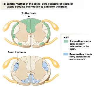
White matter consists of tracts of axons carrying information to and from the brain.
Ascending Tracts: Carry sensory information to the brain (dorsal and external lateral).
Descending Tracts: Carry motor commands from the brain to motor neurons (ventral and interior lateral).
Spinal Reflexes
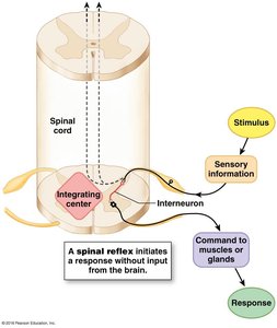
The spinal cord can act as an integrating center, initiating responses to stimuli without input from the brain. This is essential for rapid, automatic actions.
Reflex Arc: Involves sensory input, integration in the spinal cord, and motor output.
Importance: Critical for body movement and protection.
Brain Structure and Divisions
The brain is the organ responsible for complex behaviors and higher functions. It contains approximately 85 billion neurons and is divided into six major regions.
Cerebrum
Cerebellum
Diencephalon
Midbrain
Pons
Medulla
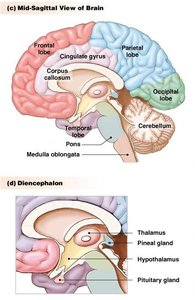
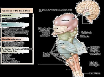
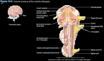
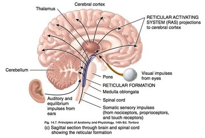
Brainstem: Medulla, Pons, Midbrain, Reticular Formation
The brainstem is the oldest and most primitive region of the brain, containing ascending and descending tracts, nuclei, and most cranial nerves.
Medulla: Controls involuntary functions (cardiovascular, respiratory centers), contains the pyramids where corticospinal tracts decussate, and centers for vomiting, swallowing, coughing, sneezing, and hiccupping.
Pons: Relays information between cerebellum and cerebrum, assists medulla in breathing coordination.
Midbrain: Controls eye movement, relays auditory and visual reflexes, contains substantia nigra.
Reticular Formation: Regulates consciousness, arousal, attention, muscle tone, and vital functions.
Cerebellum
The cerebellum is the second largest brain structure, responsible for processing sensory information related to movement and coordinating its execution.
Functions: Regulates posture, balance, and smooths movements by sending feedback to motor areas of the cerebral cortex.
Diencephalon: Thalamus and Hypothalamus
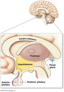
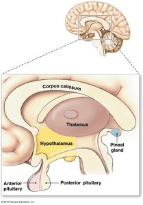
The diencephalon lies between the brainstem and cerebrum and includes the thalamus, hypothalamus, pineal gland, and pituitary gland.
Thalamus: Acts as a relay center for sensory information, sending it to relevant cortical areas.
Pineal Gland: Releases melatonin, regulating sleep/wake cycles.
Hypothalamus: Center for homeostasis, influences autonomic and endocrine functions.
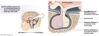
Pituitary Gland: Divided into anterior (endocrine tissue) and posterior (neural tissue) lobes, controls growth, metabolism, and reproduction.
Pituitary Gland: Structure and Function
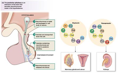
Posterior Pituitary (Neurohypophysis): Extension of the brain, releases neurohormones produced in the hypothalamus.
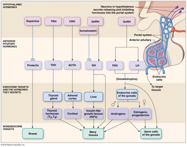
Anterior Pituitary: True endocrine gland, releases hormones that regulate other endocrine organs.
Hypothalamic-Hypophyseal Portal System: Connects hypothalamus and anterior pituitary, allowing hormone transport.
Summary Table: CNS Divisions and Functions
Region | Main Function | Key Structures |
|---|---|---|
Spinal Cord | Information relay, reflexes | Gray/white matter, dorsal/ventral roots |
Brainstem | Basic life functions, relay | Medulla, pons, midbrain, reticular formation |
Cerebellum | Movement coordination | Cerebellar hemispheres |
Diencephalon | Sensory relay, homeostasis | Thalamus, hypothalamus, pineal, pituitary |
Key Terms and Definitions
Nucleus: Cluster of cell bodies in the CNS.
Ganglion: Cluster of cell bodies in the PNS.
Afferent: Sensory input to CNS.
Efferent: Motor output from CNS.
Decussation: Crossing over of nerve fibers (e.g., in medulla pyramids).
Additional info: Academic context was added to clarify the functions and relationships of CNS structures, as well as to provide definitions and a summary table for exam preparation.
