 뒤로
Anatomy of the Respiratory System: Lab Study Guide
스터디 가이드 - 스마트 노트
 자료에 맞춘 맞춤형 노트, 핵심 정의, 예시, 맥락을 확장해 제공합니다.
Respiratory System Anatomy
Overview of the Respiratory System
The respiratory system is responsible for the exchange of gases (oxygen and carbon dioxide) between the body and the environment. It consists of a series of organs and structures that conduct air to the lungs and facilitate gas exchange at the alveolar level.
Major Functions: Air conduction, gas exchange, sound production, olfaction, and protection against pathogens.
Main Divisions: Upper respiratory tract (nose, nasal cavity, pharynx, larynx) and lower respiratory tract (trachea, bronchi, lungs).
Upper Respiratory Tract
Nasal Cavity and Associated Structures
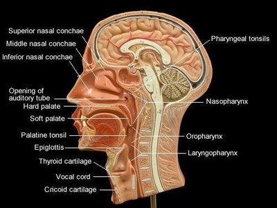
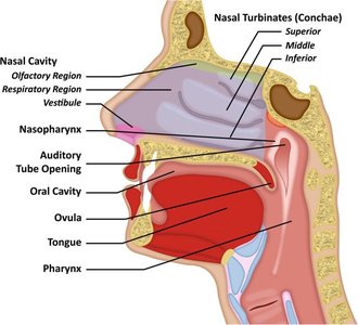
The nasal cavity is the entry point for air and is responsible for filtering, warming, and humidifying incoming air. It is divided by the nasal septum and contains several important anatomical features.
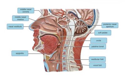
Nasal Vestibule: The anterior part of the nasal cavity just inside the nostrils (external nares).
Nasal Conchae (Turbinates): Three bony projections (superior, middle, inferior) that increase surface area and create turbulence to filter and humidify air.
Nasal Meatuses: Passages beneath each concha (superior, middle, inferior) that direct airflow.
Internal Nares (Choanae): Openings at the posterior end of the nasal cavity leading to the pharynx.
Olfactory Region: Area at the roof of the nasal cavity containing olfactory receptors for smell.
Pharynx
The pharynx is a muscular tube that serves as a passageway for both air and food. It is divided into three regions:
Nasopharynx: Located posterior to the nasal cavity; serves only as an air passage.
Oropharynx: Posterior to the oral cavity; serves as a passage for both air and food.
Laryngopharynx: Inferior portion; leads to the larynx (air) and esophagus (food).
Larynx (Voice Box)
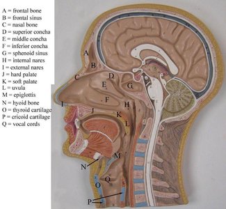
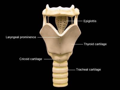
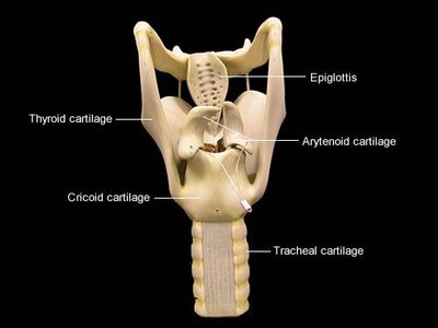
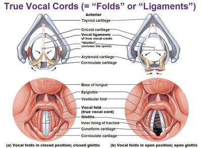
The larynx is a cartilaginous structure that connects the pharynx to the trachea and houses the vocal cords. It plays a crucial role in sound production and protecting the lower airways during swallowing.
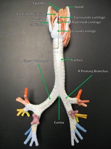
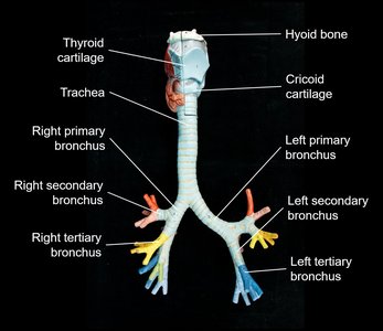
Thyroid Cartilage: Largest cartilage; forms the laryngeal prominence (Adam's apple).
Cricoid Cartilage: Ring-shaped cartilage below the thyroid cartilage.
Epiglottis: Leaf-shaped flap that covers the glottis during swallowing to prevent food from entering the airway.
Vocal Cords (Folds): Mucosal folds that vibrate to produce sound; true vocal cords are responsible for sound, while false vocal cords (vestibular folds) do not produce sound.
Lower Respiratory Tract
Trachea and Bronchial Tree
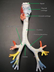
The trachea (windpipe) is a flexible tube supported by C-shaped cartilaginous rings. It conducts air from the larynx to the bronchi, which branch into the lungs.
Tracheal Cartilages: C-shaped rings that keep the trachea open.
Primary (Main) Bronchi: Right and left branches from the trachea entering each lung.
Secondary (Lobar) Bronchi: Branches that supply each lobe of the lungs (three on the right, two on the left).
Tertiary (Segmental) Bronchi: Further subdivisions supplying bronchopulmonary segments.
Bronchioles: Smallest airways lacking cartilage, leading to alveolar ducts and sacs.
Lungs and Alveoli
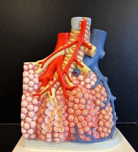
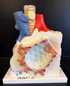
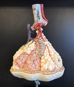
The lungs are paired organs responsible for gas exchange. Each lung is divided into lobes and contains millions of alveoli, which are the primary sites of gas exchange.
Lobes: Right lung has three lobes (superior, middle, inferior); left lung has two lobes (superior, inferior) and a cardiac notch.
Alveoli: Tiny air sacs surrounded by capillaries where oxygen and carbon dioxide are exchanged.
Pulmonary Capillaries: Network of blood vessels surrounding alveoli for efficient gas exchange.
Pulmonary Artery: Brings deoxygenated blood from the heart to the lungs.
Pulmonary Vein: Returns oxygenated blood from the lungs to the heart.
Histology of the Respiratory System
Tracheal Wall Structure
The tracheal wall is composed of several layers, each with distinct functions:
Mucosa: Innermost layer with pseudostratified ciliated columnar epithelium and goblet cells that secrete mucus.
Submucosa: Connective tissue containing seromucous glands.
Cartilage: Hyaline cartilage rings providing structural support.
Adventitia: Outermost connective tissue layer anchoring the trachea.
Layer | Main Features |
|---|---|
Mucosa | Pseudostratified ciliated epithelium, goblet cells |
Submucosa | Seromucous glands |
Cartilage | Hyaline cartilage rings |
Adventitia | Connective tissue |
Lung Histology
The lungs contain a variety of structures visible under the microscope:
Bronchi: Larger airways with cartilage plates in their walls.
Bronchioles: Smaller airways lacking cartilage, lined by simple cuboidal or columnar epithelium.
Alveoli: Lined by simple squamous epithelium (type I pneumocytes) and scattered type II pneumocytes (produce surfactant).
Alveolar Macrophages: Immune cells that remove debris and pathogens from alveolar spaces.
Structure | Histological Features |
|---|---|
Bronchi | Cartilage plates, pseudostratified ciliated epithelium |
Bronchioles | No cartilage, simple cuboidal/columnar epithelium |
Alveoli | Simple squamous epithelium, thin walls for gas exchange |
Summary Table: Key Respiratory Structures
Structure | Function |
|---|---|
Nasal Cavity | Filters, warms, humidifies air |
Pharynx | Passage for air and food |
Larynx | Sound production, airway protection |
Trachea | Conducts air to bronchi |
Bronchi | Distribute air to lungs |
Bronchioles | Control airflow, lead to alveoli |
Alveoli | Site of gas exchange |
Additional info:
Respiratory system histology is essential for understanding disease processes such as asthma, chronic bronchitis, and emphysema.
Surfactant produced by type II alveolar cells reduces surface tension, preventing alveolar collapse.
