 뒤로
LEC 5
스터디 가이드 - 스마트 노트
 자료에 맞춘 맞춤형 노트, 핵심 정의, 예시, 맥락을 확장해 제공합니다.
Appendicular Muscles: Muscles Positioning the Pectoral Girdle
Overview of Muscles Positioning the Pectoral Girdle
The pectoral girdle is stabilized and moved by several muscles that originate from the axial skeleton and insert onto the scapula or clavicle. These muscles are essential for shoulder movement and upper limb function.
Levator scapulae: Elevates the scapula.
Pectoralis minor: Depresses and protracts the scapula.
Rhomboid major/minor: Adduct and downwardly rotate the scapula.
Serratus anterior: Protracts the scapula and holds it against the thoracic wall.
Subclavius: Depresses and protracts the clavicle.
Trapezius: Elevates, retracts, depresses, and rotates the scapula.
Example: The trapezius muscle is involved in shrugging the shoulders.
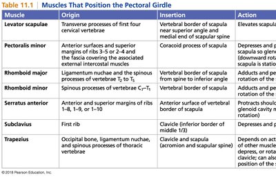
Muscle | Origin | Insertion | Action |
|---|---|---|---|
Levator scapulae | Transverse processes of first four cervical vertebrae | Vertebral border of scapula near superior angle and medial end of scapular spine | Elevates scapula |
Pectoralis minor | Anterior surfaces and superior margins of ribs 3–5 or 2–4 and the fascia covering the associated external intercostal muscles | Coracoid process of scapula | Depresses and protracts scapula; rotates scapula downward; elevates ribs if scapula is stationary |
Rhomboid major | Ligamentum nuchae and the spinous processes of vertebrae T2 to T5 | Vertebral border of scapula from spine to inferior angle | Adducts scapula and performs downward rotation |
Rhomboid minor | Spinous processes of vertebrae C7–T1 | Vertebral border of scapula | Adducts scapula and performs downward rotation |
Serratus anterior | Anterior and superior margins of ribs 1–8, 1–9, or 1–10 | Anterior surface of vertebral border of scapula | Protracts scapula; upward rotation |
Subclavius | First rib | Clavicle (inferior border of middle 1/3) | Depresses and protracts clavicle |
Trapezius | Occipital bone, ligamentum nuchae, and spinous processes of thoracic vertebrae | Clavicle and scapula (acromion and scapular spine) | Depends on active region; may elevate, retract, depress, or rotate scapula upward; extend neck |
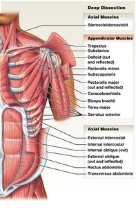
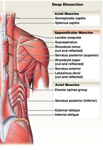
Anatomical Dissection of Pectoral Girdle Muscles
Deep and superficial dissections reveal the arrangement of axial and appendicular muscles around the shoulder. Understanding their locations helps in identifying their functions and clinical relevance.
Appendicular muscles include trapezius, subclavius, deltoid, pectoralis major/minor, supraspinatus, infraspinatus, teres major/minor, serratus anterior, and others.
Axial muscles include sternocleidomastoid, intercostals, obliques, rectus abdominis, and transversus abdominis.
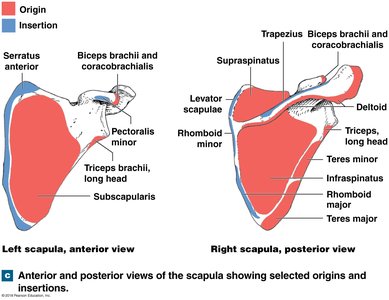
Origins and Insertions on the Scapula
The scapula serves as a major site for muscle attachment, with different muscles originating or inserting on its anterior and posterior surfaces. This arrangement allows for complex movements of the shoulder and arm.
Anterior view: Serratus anterior, biceps brachii, pectoralis minor, triceps brachii, subscapularis.
Posterior view: Trapezius, supraspinatus, infraspinatus, teres major/minor, rhomboid major/minor, deltoid.
Appendicular Muscles: Muscles Moving the Arm
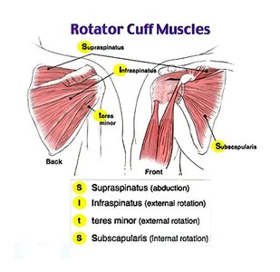
Rotator Cuff Muscles
The rotator cuff is a group of four muscles that stabilize the shoulder joint and allow for a range of movements. These muscles originate from the scapula and insert onto the humerus.
Supraspinatus: Abduction of the arm.
Infraspinatus: External rotation of the arm.
Teres minor: External rotation of the arm.
Subscapularis: Internal rotation of the arm.
Example: Injury to the rotator cuff can cause pain and limit shoulder movement.
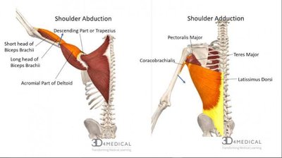
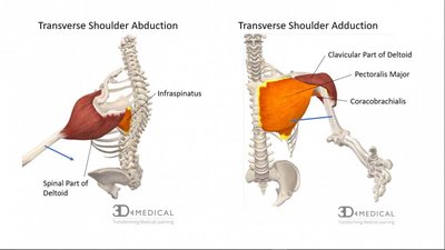
Shoulder Abduction and Transverse Shoulder Abduction
Shoulder abduction is the movement of the arm away from the body, primarily performed by the deltoid and assisted by other muscles. Transverse shoulder abduction involves moving the arm horizontally away from the midline.
Muscles involved: Deltoid, trapezius, biceps brachii, pectoralis major, coracobrachialis, teres major, latissimus dorsi, infraspinatus.
Different parts of the deltoid and other muscles contribute to these movements.
Appendicular Muscles: Muscles of the Upper Arm
Major Muscles of the Upper Arm
The upper arm contains muscles that move the forearm at the elbow. These include the biceps brachii, triceps brachii, and brachialis, each with distinct actions.
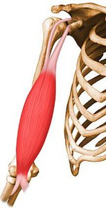
Biceps brachii: Flexes the elbow and supinates the forearm.
Triceps brachii: Extends the elbow.
Brachialis: Flexes the elbow.
Example: The biceps brachii is commonly used in lifting objects.
Connective Tissue Framework and Compartments
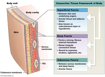
Connective Tissue Framework of the Body
Connective tissue forms the internal framework of the body, connecting organs and providing structural support. Fascia is a key component, with superficial, deep, and subserous layers.
Superficial fascia: Between skin and underlying organs; contains areolar and adipose tissue.
Deep fascia: Strong, fibrous internal framework; dense irregular connective tissue.
Subserous fascia: Between serous membranes and deep fascia; areolar tissue.
Compartments and Sectional Anatomy of the Upper Arm
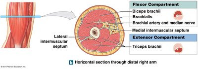
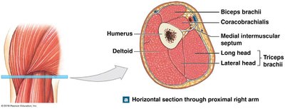
Muscles in the upper arm are organized into compartments by connective tissue septa. This arrangement allows for independent movement and clinical identification of muscle groups.
Anterior compartment: Flexor compartment (biceps brachii, brachialis).
Posterior compartment: Extensor compartment (triceps brachii).
Intermuscular septa separate these compartments.
Appendicular Muscles: Muscles that Move the Thigh
Muscle Groups Moving the Thigh
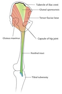
Muscles that move the thigh are grouped based on their location and function. They attach to the ilium, femur, or iliotibial tract and are responsible for movements such as abduction, adduction, and rotation.
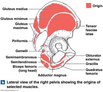
Gluteal group: Gluteus maximus, medius, minimus, tensor fasciae latae.
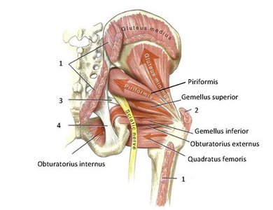
Lateral rotator group: Obturator muscles, piriformis, gemelli, quadratus femoris.
Adductor group: Adductor brevis, longus, magnus, pectineus, gracilis.
Iliopsoas group: Iliacus, psoas major.
Functional Actions of Thigh Muscles
Gluteal muscles cover the lateral surface of the ilium and are involved in abduction and rotation. Lateral rotators move the femur laterally, while adductors pull the leg toward the midline. Most adductors insert on the linea aspera of the femur.
Abduction: Performed by gluteus medius and minimus.
Lateral rotation: Performed by piriformis, gemelli, obturator muscles.
Adduction: Performed by adductor group.
Example: A "pulled groin" is an injury to the adductor muscles.

Appendicular Muscles: Muscles that Move the Leg
Quadriceps and Hamstrings
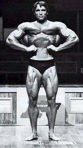
The quadriceps femoris group are extensors of the knee, while the hamstrings are flexors. The sartorius is the longest muscle in the body, running from the hip to the knee.
Quadriceps femoris: Vastus intermedius, lateralis, medialis, rectus femoris.
Hamstrings: Biceps femoris, semimembranosus, semitendinosus.
Sartorius: Longest muscle, not part of quads.
Popliteus: Unlocks the knee joint.
Compartments and Sectional Anatomy of the Thigh
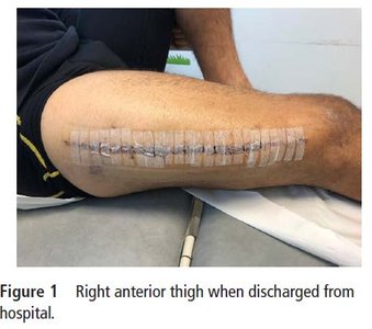
The thigh is divided into anterior, posterior, and medial (adductor) compartments by intermuscular septa. Trauma to these compartments can cause compartment syndrome, a serious condition where pressure reduces blood flow and damages tissue.
Acute compartment syndrome: Emergency, can cause permanent damage.
Chronic compartment syndrome: Painful, often due to intense activity.
Cardiac Muscle and the Cardiovascular System
Structure and Function of Cardiac Muscle
Cardiac muscle is striated and contains intercalated discs, which allow for synchronized contraction through gap junctions. This creates a functional syncytium, enabling efficient heart function.
Intercalated discs: Specialized connections between cardiomyocytes.
Gap junctions: Allow ion flow and electrical coupling.
Desmosomes: Provide mechanical strength.

Heart Anatomy and Circulation
The heart is rotated slightly to the left and has four chambers, each identified by external sulci. The aorta and pulmonary trunk are major vessels, and the auricles are ear-like projections of the atria.
Aorta: Ascending, arch, descending regions.
Pulmonary trunk: Carries blood to the lungs.
Auricles: Projections of the atria.
Heart Valves
The heart contains four valves: two atrioventricular (AV) valves and two semilunar valves. The AV valves are the tricuspid (right) and bicuspid (left), while the semilunar valves are the aortic and pulmonary valves.
Tricuspid valve: Right AV valve.
Bicuspid (mitral) valve: Left AV valve.
Aortic valve: Semilunar valve.
Pulmonary valve: Semilunar valve.
Pericardium and Pericardial Cavity
The heart is surrounded by the pericardium, which consists of an outer fibrous layer and an inner serous layer. The serous pericardium has visceral and parietal layers, with the pericardial cavity containing fluid to reduce friction.
Fibrous pericardium: Outer layer.
Serous pericardium: Inner layer (visceral and parietal).
Pericardial cavity: Contains pericardial fluid.
Conducting System of the Heart
The heart's rhythm is controlled by pacemaker cells and the conducting system, which ensures proper contraction sequence. Electrocardiograms (ECGs) record the electrical activity of the heart.
Pacemaker cells: Generate automatic rhythm.
ECG: Measures electrical signals.
Coronary Blood Vessels
Coronary arteries supply blood to the heart muscle, with the left coronary artery having more branches than the right. Coronary veins drain blood into the coronary sinus or directly into the right atrium.
Right coronary artery: Circles heart to the right.
Left coronary artery: Circles heart to the left, more branches.
Coronary sinus: Main venous drainage.
Autonomic Control of the Heart
The heart rate is set by the SA node but can be modified by the autonomic nervous system. Sympathetic stimulation increases heart rate and force, while parasympathetic stimulation decreases them.
Norepinephrine: Increases heart rate (sympathetic).
Acetylcholine: Decreases heart rate (parasympathetic).
Cardiac centers: Located in the medulla oblongata.
