 뒤로
LEC 3
스터디 가이드 - 스마트 노트
 자료에 맞춘 맞춤형 노트, 핵심 정의, 예시, 맥락을 확장해 제공합니다.
Appendicular Skeleton
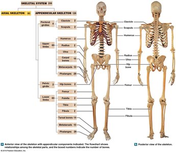
Overview of the Appendicular Skeleton
The appendicular skeleton consists of all bones that are attached to the axial skeleton, including the limbs and girdles. It is essential for movement and interaction with the environment, providing attachment points for muscles and facilitating locomotion.
Components: Clavicle, scapula, humerus, radius, ulna, carpals, metacarpals, phalanges, pelvic bones, femur, patella, tibia, fibula, tarsals, metatarsals, and phalanges.
Function: Positions the limbs, supports movement, and provides sites for muscle attachment.
Difference from Axial Skeleton: No foramina in appendicular bones; nerves and vessels travel adjacent to them.
Pectoral Girdle
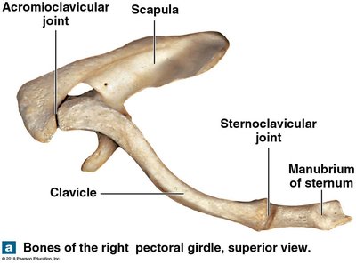
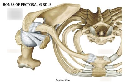
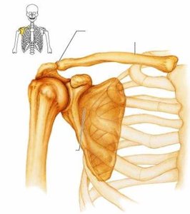
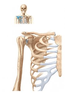
The pectoral girdle anchors the upper limb to the axial skeleton and consists of the clavicle and scapula. It provides a base for arm movement and is a key site for muscle attachment.
Clavicle: S-shaped bone connecting the sternum to the scapula.
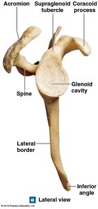
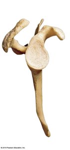
Scapula: Triangular bone with important landmarks such as the spine, acromion, coracoid process, and glenoid cavity.
Joints: Acromioclavicular and sternoclavicular joints allow for mobility and stability.
Scapula: Landmarks and Muscle Attachments
The scapula is a triangular bone with several important features for muscle attachment and articulation.
Spine: Divides the scapula into supraspinous and infraspinous fossae.
Acromion: Extends anteriorly for muscle attachment.
Glenoid cavity: Articulates with the head of the humerus.
Coracoid process: Provides additional muscle attachment.
Fossae: Supraspinous, infraspinous, and subscapular fossae are sites for muscle attachment.
Upper Limb Bones
Humerus, Radius, and Ulna
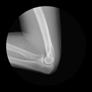
The upper limb consists of the humerus, radius, and ulna, which articulate at the elbow and wrist.
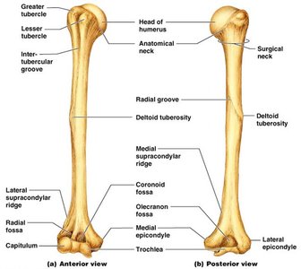
Humerus: Largest bone of the upper limb, articulates with scapula at the shoulder and with radius and ulna at the elbow.
Radius: Lateral bone of the forearm, important for wrist movement.
Ulna: Medial bone of the forearm, forms the elbow joint with the humerus.
Landmarks: Tuberosities, epicondyles, and fossae are important for muscle attachment and articulation.
Hand Bones
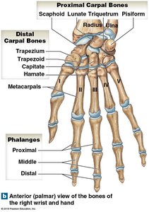
The hand is composed of carpals, metacarpals, and phalanges, allowing for complex movements and dexterity.
Carpals: Eight wrist bones arranged in two rows.
Metacarpals: Five bones forming the palm, numbered I-V.
Phalanges: Fourteen bones in each hand; thumb (pollex) has two, others have three.
Pelvic Girdle and Lower Limb
Pelvic Girdle Structure
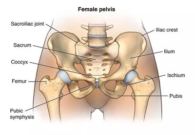
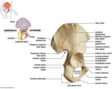
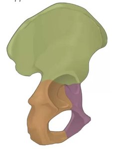
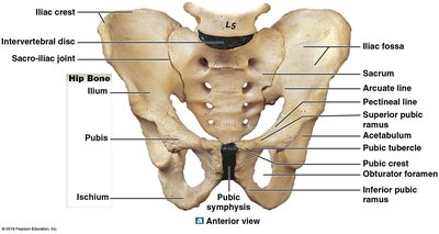
The pelvic girdle connects the lower limb to the axial skeleton and is composed of two hip bones, each formed by the fusion of the ilium, ischium, and pubis.
Acetabulum: Socket for articulation with the femur.
Pubic symphysis: Fibrous cartilage pad connecting the pubic bones.
Landmarks: Iliac crest, sciatic notch, obturator foramen, and arcuate line.
Femur and Lower Limb Bones
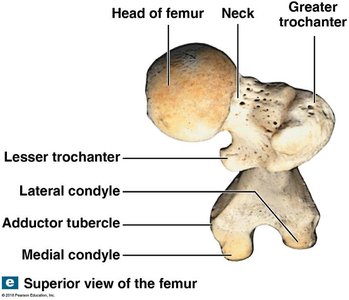
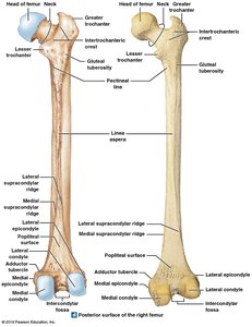
The femur is the largest bone in the body, articulating with the pelvis at the acetabulum and with the tibia and patella at the knee.
Femur: Head, neck, greater and lesser trochanters, condyles, and epicondyles are key landmarks.
Tibia: Main weight-bearing bone of the leg.
Fibula: Lateral bone of the leg, important for muscle attachment.
Patella: Sesamoid bone protecting the knee joint.
Foot Bones
The foot is composed of tarsals, metatarsals, and phalanges, designed for support and mobility.
Tarsals: Seven ankle bones, including talus and calcaneus.
Metatarsals: Five bones forming the arch of the foot.
Phalanges: Fourteen bones; big toe (hallux) has two, others have three.
Joints (Articulations)
Classification of Joints
Joints are classified based on their function and range of motion.
Synarthrosis: Immovable joints (e.g., sutures in the skull).
Amphiarthrosis: Slightly movable joints (e.g., syndesmosis between radius and ulna, symphysis between pubic bones).
Diarthrosis: Freely movable joints, also called synovial joints (e.g., shoulder, elbow, hip, knee).
Types of Synovial Joints
Synovial joints are characterized by a joint cavity, articular cartilage, and supporting ligaments.
Shoulder Joint: Ball-and-socket joint allowing wide range of motion.
Elbow Joint: Hinge joint allowing flexion and extension.
Hip Joint: Ball-and-socket joint providing stability and movement.
Knee Joint: Complex hinge joint with menisci and supporting ligaments.
Ankle Joint: Talocrural joint for dorsiflexion and plantarflexion.
Movements at Joints
Joints allow for various types of movement, classified as angular, rotational, or special.
Angular Motion: Abduction, adduction, flexion, extension, hyperextension, circumduction.
Rotational Motion: Medial and lateral rotation, pronation, supination.
Special Movements: Unique to certain joints (e.g., opposition, reposition).
Key Ligaments and Structures
Ligaments stabilize joints and connect bone to bone.
Shoulder: Glenohumeral, coracohumeral, coraco-acromial, acromioclavicular, coracoclavicular, transverse humeral ligaments.
Elbow: Radial collateral, ulnar collateral, annular ligaments.
Hip: Iliofemoral, pubofemoral, ischiofemoral, transverse acetabular ligaments.
Knee: Medial and lateral menisci, fat pads, extracapsular and intracapsular ligaments.
Ankle: Tibiotalar, fibulotalar, distal tibiofibular joints.
Summary Table: Major Bones of the Appendicular Skeleton
Region | Main Bones | Key Landmarks |
|---|---|---|
Pectoral Girdle | Clavicle, Scapula | Acromion, coracoid process, glenoid cavity |
Upper Limb | Humerus, Radius, Ulna | Epicondyles, tuberosities, olecranon |
Hand | Carpals, Metacarpals, Phalanges | Proximal/distal rows, pollex |
Pelvic Girdle | Ilium, Ischium, Pubis | Acetabulum, iliac crest, pubic symphysis |
Lower Limb | Femur, Patella, Tibia, Fibula | Trochanters, condyles, malleoli |
Foot | Tarsals, Metatarsals, Phalanges | Talus, calcaneus, hallux |
Additional info:
Mnemonic devices can help remember the order of carpal and tarsal bones.
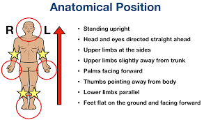
Understanding anatomical position is crucial for identifying medial and lateral structures.
Ligaments and fossae are named based on their location and function.
