 뒤로
뒤로Appendicular Skeleton: Structure and Function
스터디 가이드 - 스마트 노트
 자료에 맞춘 맞춤형 노트, 핵심 정의, 예시, 맥락을 확장해 제공합니다.
The Appendicular Skeleton
Overview
The appendicular skeleton consists of the bones of the limbs and their girdles, which attach the limbs to the axial skeleton. It is divided into the pectoral (shoulder) girdle and the pelvic (hip) girdle, each providing support and mobility for the upper and lower limbs, respectively.
Pectoral girdle: Attaches upper limbs to the trunk.
Pelvic girdle: Attaches lower limbs to the trunk.
The Pectoral Girdle
Structure and Function
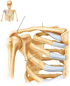
The pectoral girdle is composed of the clavicles (anteriorly) and scapulae (posteriorly). It provides attachment sites for muscles that move the upper limbs and offers a high degree of mobility due to the shallow socket of the shoulder joint and the lack of direct attachment of the scapulae to the axial skeleton.
Clavicle: S-shaped bone, articulates medially with the sternum and laterally with the scapula. Acts as a brace for the scapula and arms.
Scapula: Thin, triangular bone with three borders (superior, medial, lateral) and three angles (superior, lateral, inferior). Features include the spine, acromion, coracoid process, and glenoid cavity.
The Upper Limb
Arm, Forearm, and Hand
The upper limb consists of 30 bones forming the arm, forearm, and hand. The arm contains the humerus, the forearm contains the radius and ulna, and the hand contains carpal, metacarpal, and phalangeal bones.
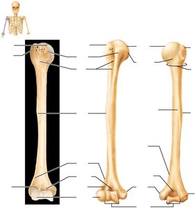
Humerus: Largest bone of the upper limb, articulates with the scapula and forearm bones. Features include the head, anatomical neck, greater and lesser tubercles, surgical neck, deltoid tuberosity, and epicondyles.
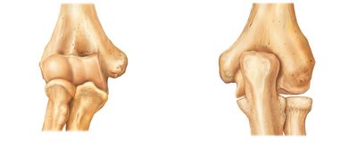
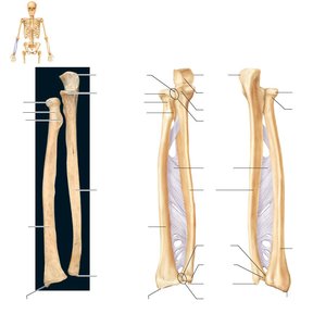
Radius and Ulna: Parallel bones of the forearm, connected by an interosseous membrane. The ulna forms the major portion of the elbow joint, while the radius is lateral and articulates with the wrist.
Hand: Composed of the carpus (wrist, 8 bones), metacarpus (palm, 5 bones), and phalanges (fingers, 14 bones).
The Pelvic Girdle
Structure and Function
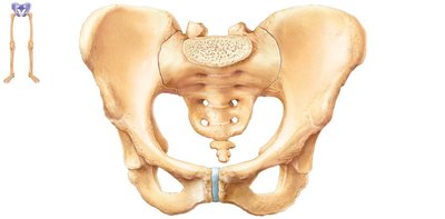
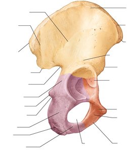
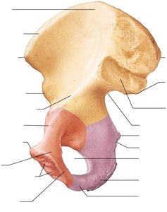
The pelvic girdle, or hip girdle, is formed by two hip bones (coxal bones) and the sacrum. It attaches the lower limbs to the axial skeleton, transmits weight, and supports pelvic organs. The coxal bone is formed by the fusion of the ilium, ischium, and pubis, with the acetabulum serving as the socket for the femur.
Ilium: Superior region, articulates with the sacrum.
Ischium: Posteroinferior region, features include the ischial spine and tuberosity.
Pubis: Anterior region, forms the pubic symphysis and pubic arch.
Pelvic Structure and Childbearing
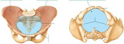
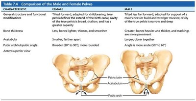
The pelvis is adapted for childbearing in females, being wider, shallower, and lighter. The pelvic brim defines the pelvic inlet, and the true pelvis forms the birth canal.
Comparison of Male and Female Pelves
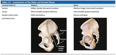
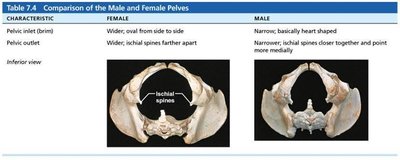
Male and female pelves differ in structure and function, with the female pelvis adapted for childbirth.
Characteristic | Female | Male |
|---|---|---|
General structure | Wider, lighter, rounder | Narrower, heavier, more acute angle |
Bone thickness | Lighter, thinner | Heavier, thicker |
Acetabula | Smaller, farther apart | Larger, closer together |
Pubic arch/angle | Broader (80–90°) | More acute (50–60°) |
Sacrum | Wider, shorter | Narrower, longer |
Coccyx | More movable, projects inferiorly | Less movable, projects anteriorly |
Pelvic inlet | Wider, oval | Narrow, heart-shaped |
Pelvic outlet | Wider | Narrower |
The Lower Limb
Thigh, Leg, and Foot
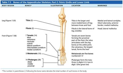
The lower limb supports the weight of the body and is subjected to significant forces. It consists of the thigh (femur), leg (tibia and fibula), and foot (tarsals, metatarsals, phalanges).
Femur: Largest and strongest bone, articulates with the hip and knee. Features include the head, neck, trochanters, condyles, epicondyles, and patellar surface.
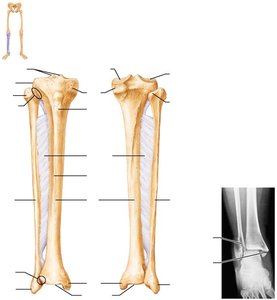
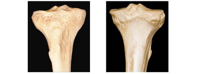
Tibia: Medial bone, weight-bearing, articulates with femur and foot.
Fibula: Lateral bone, not weight-bearing, provides muscle attachment.
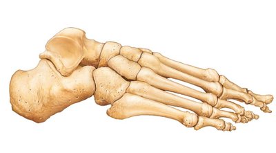
Foot: Includes tarsals (7 bones), metatarsals (5 bones), and phalanges (14 bones).
Arches of the Foot
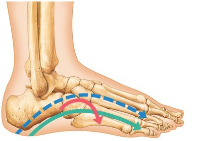
The foot has three arches maintained by bones, ligaments, and tendons, allowing it to bear weight and absorb shock.
Lateral longitudinal arch: Low curve, elevates lateral part of foot.
Medial longitudinal arch: Higher curve, elevates medial part.
Transverse arch: Runs obliquely across the foot.
Developmental Aspects of the Skeleton
Infant Skull and Spine
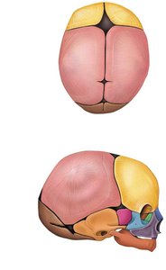
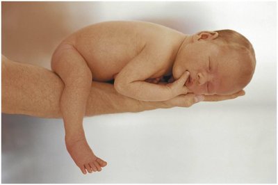
Infant skulls have more bones than adult skulls, with unfused bones and fontanelles that allow for brain growth and ease of birth. The spine of a newborn is C-shaped, with primary curvatures in the thorax and sacrum, and secondary curvatures developing in the cervical and lumbar regions as the child grows.
Key Terms and Concepts
Acetabulum: Deep socket in the hip bone that receives the head of the femur.
Fontanelles: Soft spots in the infant skull where bones are not yet fused.
Interosseous membrane: Fibrous sheet connecting parallel bones (radius/ulna, tibia/fibula).
Pelvic brim: Continuous oval ridge defining the pelvic inlet.
Trochanter: Large, blunt, irregularly shaped process on the femur for muscle attachment.
Example: Application in Clinical Practice
Understanding the differences between male and female pelves is crucial in obstetrics for assessing childbirth risks. Knowledge of bone features aids in diagnosing fractures and planning orthopedic interventions.
Additional info: The notes expand on brief points with academic context, definitions, and examples to ensure completeness and clarity for exam preparation.
