 뒤로
Appendicular Skeleton: Structure, Function, and Key Joints
스터디 가이드 - 스마트 노트
 자료에 맞춘 맞춤형 노트, 핵심 정의, 예시, 맥락을 확장해 제공합니다.
Appendicular Skeleton Overview
Divisions of the Skeletal System
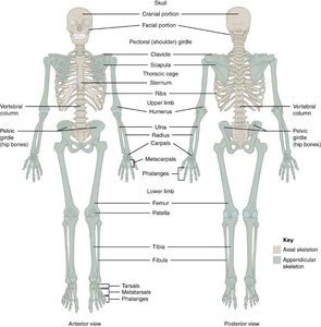
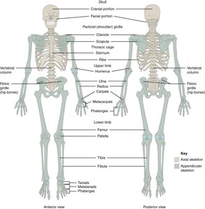
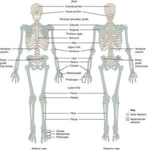
The human skeleton is divided into two main parts: the axial skeleton and the appendicular skeleton. The appendicular skeleton consists of 126 bones and is primarily responsible for movement and manipulation of the environment.
Axial Skeleton: Composed of 80 bones, protects vital organs such as the brain, spinal cord, heart, and lungs.
Appendicular Skeleton: Includes bones of the upper and lower limbs, pectoral girdle, and pelvic girdle. It facilitates locomotion and manipulation.
Components of the Appendicular Skeleton
Upper Limbs: Pectoral girdle, upper arm (humerus), forearm (radius and ulna), and hand (carpals, metacarpals, phalanges).
Lower Limbs: Pelvic girdle, upper leg (femur), lower leg (tibia and fibula), and foot (tarsals, metatarsals, phalanges).
Pectoral (Shoulder) Girdle
Structure and Function
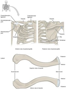
The pectoral girdle attaches the upper limbs to the axial skeleton and allows for extensive mobility. It consists of the clavicle and scapula, stabilized by muscles and ligaments.
Clavicle: S-shaped bone on the anterior side of the shoulder.
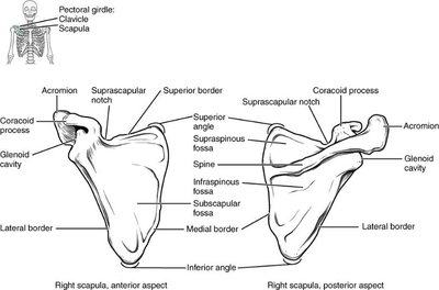
Scapula: Flat, triangular bone on the posterior side, attached to ribs by muscles and tendons.
Joints: Sternoclavicular, acromioclavicular, and glenohumeral joints provide mobility and stability.
Glenohumeral (Shoulder) Joint
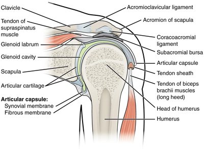
The glenohumeral joint is a ball-and-socket joint with the largest range of motion in the body.
Articulation: Head of humerus fits into the shallow glenoid cavity of the scapula.
Glenoid Labrum: Fibrocartilage deepens the cavity for stability.
Support: Ligaments, muscles (especially rotator cuff), and bursae provide structural support and reduce friction.
Bones and Joints of the Upper Limb
Major Bones
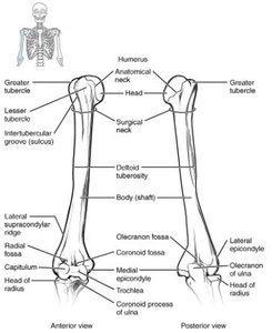
Humerus: Long bone of the upper arm.
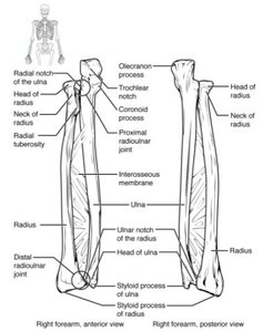
Radius: Located on the thumb side of the forearm.
Ulna: Located on the pinky side of the forearm.
Key Joints
Glenohumeral Joint: Allows wide range of motion.
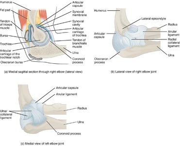
Proximal Radioulnar Joint (Elbow): Allows forearm rotation.
Distal Radioulnar Joint (Wrist): Allows pronation and supination.
Interosseous Membrane: Maintains forearm integrity.
Muscles of the Upper Limb
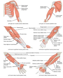
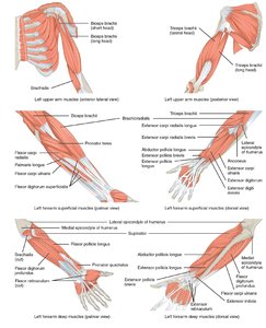
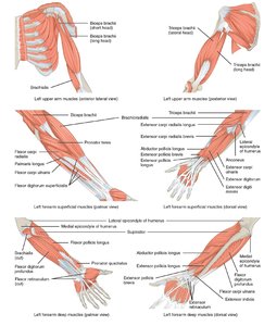
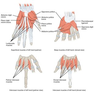
Muscles that move the arm cross the shoulder joint and are classified by their location and function.
Flexion: Biceps brachii, brachialis, brachioradialis.
Extension: Triceps brachii, anconeus.
Pronation: Pronator teres, pronator quadratus.
Supination: Supinator.
Bones and Joints of the Lower Limb
Pelvic (Hip) Girdle
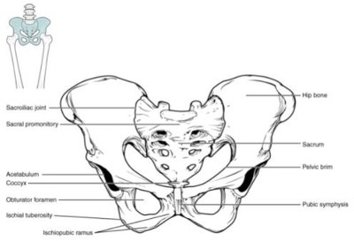
The pelvic girdle provides a stable base for the lower limbs and supports the weight of the upper body.
Structure: Consists of the sacrum and two coxal bones (each fused from ilium, ischium, and pubis).
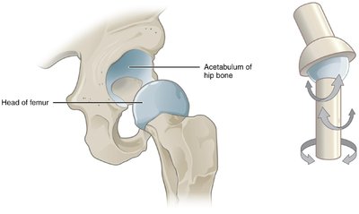
Acetabulum: Deep socket for the head of the femur, providing stability.
Function: Weight-bearing, transmits weight, supports pelvic organs.
Bones of the Lower Limb
Femur: Longest and heaviest bone in the body.
Tibia: Larger, weight-bearing bone of the lower leg.
Fibula: Smaller, stabilizing bone of the lower leg.
Patella: Largest sesamoid bone, stabilizes knee joint.
Knee Joint
The knee joint is a hinge joint formed by the distal femur, patella, and proximal tibia.
Menisci: Medial and lateral fibrocartilage discs provide padding and stability.
Ligaments: Anterior and posterior cruciate ligaments, collateral ligaments, and transverse ligament stabilize the joint.
Bones of the Foot
Tarsals: Includes talus (articulates with tibia and fibula) and calcaneus (heel bone).
Metatarsals: Form the framework of the foot.
Phalanges: Allow flexion and extension of toes.
Arches: Medial, lateral, and transverse arches distribute weight and provide leverage.
Ankle and Foot Joints
Talocrural (Ankle) Joint: Uniaxial hinge joint between talus and distal tibia/fibula, allows dorsiflexion and plantar flexion.
Subtalar Joint: Between talus and calcaneus, allows inversion and eversion.
Ligaments: Provide stability and prevent abnormal movements.
Intrinsic Muscles: Support arches and move toes.
Ossification of Appendicular Bones
Bone Development
Most girdle and limb bones develop by endochondral ossification, except the clavicle.
Endochondral Ossification: Process where cartilage is replaced by bone tissue.
Clavicle: Develops by intramembranous ossification.
Summary Table: Major Bones of the Appendicular Skeleton
Region | Main Bones | Key Joints |
|---|---|---|
Upper Limb | Clavicle, Scapula, Humerus, Radius, Ulna, Carpals, Metacarpals, Phalanges | Shoulder (glenohumeral), Elbow, Wrist |
Lower Limb | Pelvic girdle, Femur, Tibia, Fibula, Patella, Tarsals, Metatarsals, Phalanges | Hip, Knee, Ankle |
Additional info:
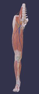
Lower limb bones are larger and stronger than upper limb bones due to their weight-bearing function.
Both the shoulder and hip joints are multiaxial ball-and-socket joints, but the hip joint is deeper and more stable.
Muscles of the lower limb are larger and more powerful to support movement and posture.
