 뒤로
Articulations: Structure, Classification, and Function of Joints
스터디 가이드 - 스마트 노트
 자료에 맞춘 맞춤형 노트, 핵심 정의, 예시, 맥락을 확장해 제공합니다.
Articulations
Introduction to Articulations
Articulations, or joints, are the locations where two or more bones meet. They play a crucial role in movement, stability, and support within the skeletal system. Joints are classified both functionally and structurally, which determines their mobility and anatomical features. 
Functional Classification of Joints
Types Based on Mobility
Joints are classified by the degree of movement they allow:
Synarthrosis: No movement between bones (e.g., sutures of the skull).
Amphiarthrosis: Small amount of movement between bones (e.g., syndesmosis between tibia and fibula).
Diarthrosis: Freely moveable joints (e.g., most synovial joints).
Example: The knee joint is a diarthrosis, allowing significant movement.
Structural Classification of Joints
Fibrous Joints
Fibrous joints are connected by dense regular collagenous connective tissue. They generally allow little to no movement.
Suture: Immovable; found between bones of the cranium.
Gomphosis: Immovable; between a tooth and its bony socket, held by the periodontal ligament.
Syndesmosis: Allows a small amount of movement; found between radius/ulna and tibia/fibula, connected by interosseous ligaments.
Cartilaginous Joints
Cartilaginous joints are held together by cartilage and allow limited movement.
Synchondrosis: Hyaline cartilage; examples include epiphyseal plates in growing bones.
Symphysis: Fibrocartilage; examples include intervertebral joints and pubic symphysis.
Synovial Joints
Synovial joints are diarthrotic and have a fluid-filled cavity between articulating bones. They are the most mobile and complex type of joint.
Each bone's articulating surface is covered with a layer of hyaline cartilage.
A joint cavity (synovial cavity) separates the bones.
The articular capsule consists of an outer fibrous layer and an inner synovial membrane, which secretes synovial fluid.
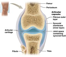
Structural Elements of Synovial Joints
Synovial Fluid
Synovial fluid is a thick liquid found within the joint cavity. It serves several functions:
Lubrication: Reduces friction between articulating surfaces.
Shock Absorption: Cushions the joint during movement.
Transport: Delivers nutrients and removes waste products from articular cartilage.
Articular Cartilage
Articular cartilage is a thin layer of hyaline cartilage covering the ends of bones in synovial joints. It reduces friction and prevents wear on the bone surfaces. 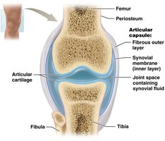
Stabilizing and Supporting Factors
Joint Stability vs. Mobility
There is an inverse relationship between joint mobility and stability: more mobility generally means less stability.
Ligament: Links bones together and provides stability.
Tendon: Connects muscle to bone, contributing to joint support.
Bursa: Synovial fluid-filled fibrous structure that minimizes friction between moving parts.
Tendon Sheath: A long bursa that surrounds and protects tendons.
Common Joint Problems
Bursitis
Bursitis is inflammation of a bursa, often caused by trauma, repetitive movements, or inflammatory diseases. Symptoms include pain and swelling.
Arthritis
Arthritis is inflammation of joints, resulting in pain and limited movement. Types include:
Osteoarthritis: Most common; caused by wear and tear, injuries, and aging.
Rheumatoid Arthritis: Autoimmune destruction of joint tissues.
Gouty Arthritis: Uric acid crystal deposits in joints.
Functional Classes of Synovial Joints
Types Based on Axes of Movement
Synovial joints are classified by the number of axes around which they move:
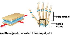
Nonaxial Joints: Motion in one or more planes without moving around an axis (e.g., intercarpals).
Uniaxial Joints: Motion around one axis (e.g., elbow).
Biaxial Joints: Motion around two axes (e.g., saddle joints).
Multiaxial (Triaxial) Joints: Motion around three axes (e.g., shoulder).
Movements at Synovial Joints
Types of Movements
Gliding Movements: Sliding motion between surfaces (nonaxial).
Angular Movements: Increase or decrease angle between bones.
Flexion: Decreases angle between bones.
Extension: Increases angle between bones.
Hyperextension: Extension beyond anatomical position.
Abduction: Moves body part away from midline.
Adduction: Moves body part toward midline.
Circumduction: Movement in a circle.
Rotation: One bone rotates around its longitudinal axis.
Special Movements: Include opposition/reposition (thumb), depression/elevation, protraction/retraction, inversion/eversion, dorsiflexion/plantarflexion, supination/pronation.
Range of Motion
Joint Range of Motion
Range of motion is the amount of movement a joint is capable of under normal circumstances. Uniaxial joints (e.g., knee) have the smallest range, while multiaxial joints (e.g., shoulder) have the greatest.
Types of Synovial Joints
Classification and Examples
Synovial joints are classified by their shape and movement:
Type | Movement | Example |
|---|---|---|
Plane Joint | Nonaxial | Intercarpal joints |
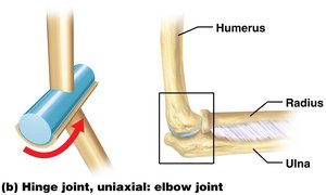
Hinge Joint | Uniaxial | Elbow joint |
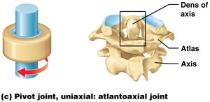
Pivot Joint | Uniaxial | Atlantoaxial joint |
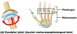
Condylar Joint | Biaxial | Metacarpophalangeal joint |
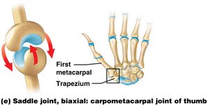
Saddle Joint | Biaxial | Carpometacarpal joint of thumb |
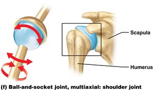
Ball-and-Socket Joint | Multiaxial | Shoulder joint |
Specific Joints
Elbow Joint
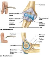
The elbow is a hinge joint composed of:
Humeroulnar joint: Trochlea of humerus and trochlear notch of ulna.
Humeroradial joint: Capitulum of humerus and head of radius.
Radial collateral ligament: Supports lateral side.
Ulnar collateral ligament: Supports medial side.
Anular ligament: Binds head of radius to neck of ulna.
Knee Joint
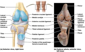
The knee is a complex hinge joint:
Tibiofemoral joint: Femoral and tibial condyles.
Patellofemoral joint: Posterior surface of patella and anterior patellar surface of femur.
Patellar ligament: Connects distal patella to anterior tibia.
Medial and lateral meniscus: C-shaped fibrocartilaginous pads between femoral and tibial condyles.
Tibial collateral ligament: Connects femur, medial meniscus, and tibia; prevents lateral shift.
ACL and PCL: Cruciate ligaments stabilizing the knee.
Shoulder Joint (Glenohumeral Joint)
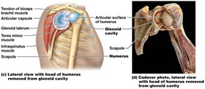
The shoulder connects the upper extremity with the axial skeleton:
Glenoid labrum: Fibrocartilaginous ring increases depth of glenoid cavity.
Biceps brachii tendon: Helps keep head of humerus within glenoid cavity.
Rotator cuff: Formed from tendons of several muscles, provides structural stabilization and strength.
Hip Joint (Coxal Joint)
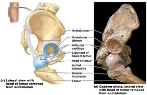
The hip is a ball-and-socket joint between the acetabulum and head of femur:
Acetabular labrum: Fibrocartilaginous ring stabilizes head of femur.
Iliofemoral ligament: Reinforces anterior joint capsule.
Ischiofemoral ligament: Spiral-shaped, supports posterior capsule.
Pubofemoral ligament: Triangular-shaped, supports inferior aspect.
Ligament of head of femur: Connects head of femur with acetabulum, provides pathway for blood vessels.
 Additional info: Synovial joints are the most common and versatile joints in the human body, allowing a wide range of movements essential for daily activities and athletic performance. Understanding their structure and function is fundamental for diagnosing and treating joint disorders.
