 뒤로
Articulations: Structure, Function, and Movements of Joints
스터디 가이드 - 스마트 노트
 자료에 맞춘 맞춤형 노트, 핵심 정의, 예시, 맥락을 확장해 제공합니다.
Articulations (Joints)
Definition and Overview
Joints, also known as articulations, are anatomical locations where two or more bones meet, or where bones interact with cartilage or teeth. The study of joints is called arthrology. Joints are classified based on their structural components and the degree of movement they allow.
Structural classification: Based on the material binding bones and the presence or absence of a joint cavity.
Functional classification: Based on the amount of movement permitted.
Structural Classification of Joints
Fibrous Joints
Fibrous joints are connected by dense regular connective tissue and lack a joint cavity. They are generally immobile or only slightly mobile. There are three main types:
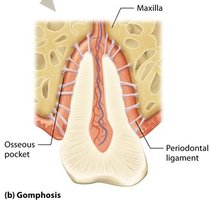
Gomphosis: Peg-in-socket joint, such as the articulation of teeth with the sockets of the mandible and maxillae. The tooth is held in place by fibrous periodontal membranes and functions as a synarthrosis (immobile joint).
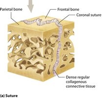
Suture: Found between skull bones, sutures are seams of very short fibers with interlocking, irregular edges. They increase strength and decrease fracture risk, allow growth in childhood, and become ossified synostoses in older adults. Functionally, they are synarthroses.
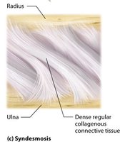
Syndesmosis: Bound by an interosseous membrane (broad ligamentous sheet), syndesmoses are found between the radius and ulna, and between the tibia and fibula. They allow slight movement (amphiarthroses) and provide a pivot for two long bones.
Cartilaginous Joints
Cartilaginous joints have either hyaline cartilage or fibrocartilage between bones and lack a joint cavity. They are either immobile or slightly mobile.
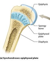
Synchondrosis: Bones joined by hyaline cartilage, such as the epiphyseal plate in growing bones and costochondral joints. These joints are immobile (synarthroses).
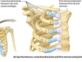
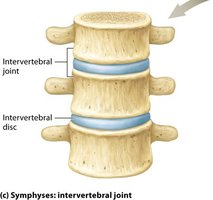
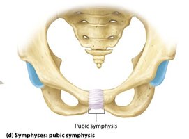
Symphysis: Pads of fibrocartilage between articulating bones, such as intervertebral joints and the pubic symphysis. These joints resist compression, act as shock absorbers, and allow slight mobility (amphiarthroses).
Synovial Joints
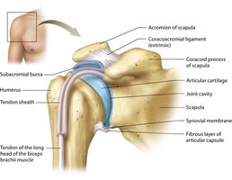
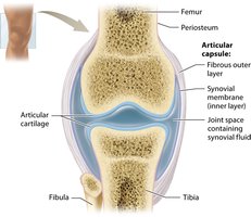
Synovial joints are characterized by bones joined by ligaments with a fluid-filled joint cavity separating bone surfaces. All synovial joints are diarthroses (freely mobile).
They have a double-layered joint capsule (articular capsule) with an outer fibrous layer and an inner synovial membrane.
Articular cartilage covers bone surfaces at the joint, reducing friction and acting as a cushion.
The joint cavity contains synovial fluid, which lubricates, nourishes, and absorbs shock.
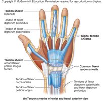
Ligaments, tendons, bursae, tendon sheaths, and fat pads are accessory structures that stabilize and protect the joint.
Functional Classification of Joints
Types Based on Movement
Synarthroses: Immobile joints (can be fibrous or cartilaginous).
Amphiarthroses: Slightly mobile joints (can be fibrous or cartilaginous).
Diarthroses: Freely mobile joints (all synovial joints).
Classification of Synovial Joints by Shape and Movement
Types of Synovial Joints
Synovial joints are further classified by the shape of their articular surfaces and the movements they allow:
Plane joint: Flat articular surfaces, uniaxial, limited gliding movement (e.g., within the hand).
Hinge joint: Convex surface within concave depression, uniaxial, like a door hinge (e.g., elbow, knee).
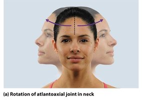
Pivot joint: Rounded surface fits into a ligament ring, uniaxial, rotation (e.g., neck).
Condylar joint: Oval, convex surface with concave surface, biaxial (e.g., fingers).
Saddle joint: Convex and concave surfaces resembling a saddle, biaxial (e.g., thumb).
Ball-and-socket joint: Spherical head fits into cuplike socket, multiaxial, most freely mobile (e.g., hip, shoulder).
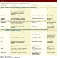
Summary Table: Structural and Functional Classification of Joints
Main Purpose: Comparison and Classification
Structural Classification | Description | Functional Classification | Examples |
|---|---|---|---|
Fibrous | Bones joined by dense connective tissue | Synarthroses (immobile), Amphiarthroses (slightly mobile) | Gomphosis (teeth), Suture (skull), Syndesmosis (radius/ulna) |
Cartilaginous | Bones joined by cartilage | Synarthroses, Amphiarthroses | Synchondrosis (epiphyseal plate, costochondral joints), Symphysis (intervertebral discs, pubic symphysis) |
Synovial | Bones joined by ligaments, joint cavity present | Diarthroses (freely mobile) | Shoulder, knee, hip, fingers, thumb |
Movements at Joints
Types of Motions
Joints allow various types of movement, classified as gliding, angular, rotational, and special movements.
Gliding: Opposing surfaces slide back-and-forth or side-to-side, limited movement (e.g., between carpals or tarsals).
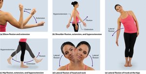
Angular: Changes the angle between bones. Includes flexion, extension, hyperextension, lateral flexion, abduction, adduction, and circumduction.
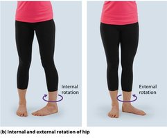
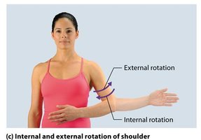
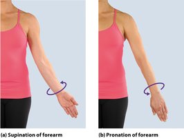
Rotational: Bone pivots on its own axis. Includes lateral and medial rotation, pronation, and supination.
Special: Unique movements such as depression, elevation, dorsiflexion, plantar flexion, inversion, eversion, protraction, retraction, opposition, and reposition.
Angular Motions
Flexion: Decreases the angle between bones (e.g., bending the elbow).
Extension: Increases the angle between bones (e.g., straightening the elbow).
Hyperextension: Extension beyond 180 degrees (e.g., looking up at the ceiling).
Lateral flexion: Trunk moves laterally in the coronal plane.
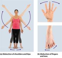
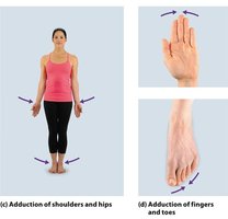
Abduction and Adduction
Abduction: Movement away from the midline (e.g., raising arms sideways).
Adduction: Movement toward the midline (e.g., lowering arms to sides).
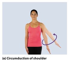
Circumduction
Circumduction: Proximal end stationary, distal end moves in a circle, forming a cone shape (e.g., drawing a circle with the arm).
Rotational Motions
Lateral rotation: Turns anterior surface of bone laterally.
Medial rotation: Turns anterior surface of bone medially.
Pronation: Medial rotation of forearm so palm faces posteriorly.
Supination: Lateral rotation of forearm so palm faces anteriorly.
Special Movements
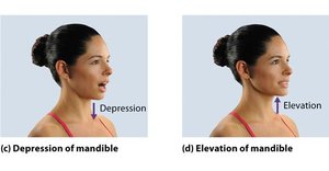
Depression: Inferior movement (e.g., opening the mouth).
Elevation: Superior movement (e.g., closing the mouth).
Special Movements of the Foot
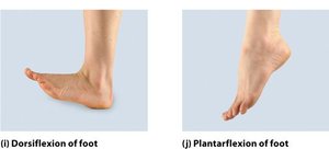
Dorsiflexion: Ankle joint bent so the dorsum of the foot moves toward the leg.
Plantar flexion: Ankle joint bent so the dorsum points inferiorly (e.g., standing on tiptoes).
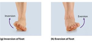
Inversion: Sole turns medially.
Eversion: Sole turns laterally.
Protraction and Retraction
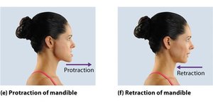
Protraction: Anterior movement from anatomic position (e.g., jutting jaw forward).
Retraction: Posterior movement from anatomic position (e.g., pulling jaw backward).
Opposition and Reposition
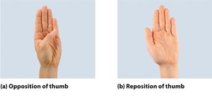
Opposition: Movement of thumb toward tips of fingers, enabling grasping.
Reposition: Moving thumb back to resting state.
Clinical View: Arthritis
Types and Symptoms
Arthritis is a group of inflammatory or degenerative diseases affecting joints, characterized by swelling, pain, and stiffness.
Gouty arthritis: Due to increased uric acid, typically affects middle-aged or older males.
Osteoarthritis: Degenerative condition due to wear of articular cartilage, common in older individuals.
Rheumatoid arthritis: Autoimmune disorder, often in younger to middle-aged women, starts with synovial membrane inflammation.
Review Questions
Given that the shoulder is a very mobile joint, how stable is it?
Are all fibrous joints synarthroses?
If a joint is a synovial joint, what else do we know about it?
Where are gomphoses located?
Functionally, how are syndesmoses classified?
Describe a suture.
What type of cartilage is found in a synchondrosis?
Functionally, how are symphyses classified?
What characteristics are shared by all synovial joints?
What functions does synovial fluid serve?
Which structural classes of synovial joint have the greatest and least mobility?
Is flexion of the elbow an angular motion or a rotational motion?
What is eversion, and where does it occur?
Standing on one's toes requires what movement?
The thumb touching another finger (on the same hand) is called what?
Additional info: Academic context was added to clarify joint types, movement classifications, and clinical relevance.
