 뒤로
Axial and Appendicular Skeleton: Bones, Markings, and Osseous Tissue
스터디 가이드 - 스마트 노트
 자료에 맞춘 맞춤형 노트, 핵심 정의, 예시, 맥락을 확장해 제공합니다.
Axial Skeleton
Overview of the Axial Skeleton
The axial skeleton forms the central axis of the human body and consists of the skull, vertebral column, and ribcage. It provides structural support, protects vital organs, and serves as attachment points for muscles.
Skull: Protects the brain and forms the structure of the face.
Vertebral column: Supports the body and protects the spinal cord.
Ribcage: Protects the heart and lungs.
Bone Markings and Features
Bone markings are anatomical features on bones where muscles, tendons, and ligaments attach, or where nerves and blood vessels pass.
Tuberosity: Large, rounded projection; may be roughened.
Crest: Narrow, prominent ridge of bone.
Trochanter: Large, blunt, irregularly shaped process (only on femur).
Line: Narrow ridge of bone; less prominent than a crest.
Tubercle: Small, rounded projection or process.
Epicondyle: Raised area on or above a condyle.
Spine: Sharp, slender, often pointed projection.
Process: Any bony prominence.
Head: Bony expansion carried on a narrow neck.
Facet: Smooth, nearly flat articular surface.
Condyle: Rounded articular projection.
Groove: Furrow.
Fissure: Narrow, slit-like opening.
Foramen: Round or oval opening through a bone.
Notch: Indentation at the edge of a structure.
Meatus: Canal-like passageway.
Sinus: Cavity within a bone, filled with air and lined with mucous membrane.
Fossa: Shallow, basin-like depression in a bone.
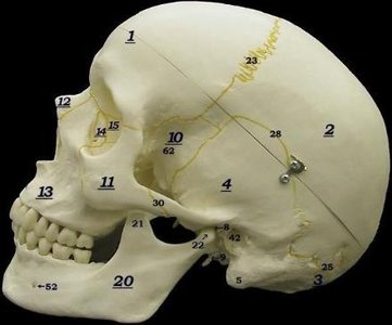
Skull Bones and Markings
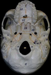
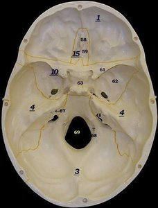
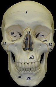
The skull is composed of several bones and features that are essential for protection and function.
Frontal bone: Forehead region.
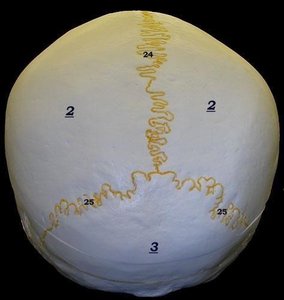
Parietal bone: Sides and roof of the cranium.
Occipital bone: Back and base of the skull.
Temporal bone: Sides and base of the skull.
Mastoid process: Prominent bone behind the ear.
Mandibular fossa: Depression for articulation with the mandible.
Styloid process: Slender pointed bone below the ear.
Sphenoid bone: Base of the skull, butterfly-shaped.
Zygomatic bone: Cheekbone.
Nasal bone: Bridge of the nose.
Maxilla: Upper jaw.
Lacrimal bone: Small bone forming part of the eye socket.
Ethmoid bone: Forms part of the nasal cavity and eye socket.
Mandible: Lower jaw.
Coronoid process: Anterior projection of the mandible.
Mandibular condyle: Posterior projection of the mandible.
Sutures: Immovable joints between skull bones (coronal, sagittal, lambdoid, squamous).
Foramen magnum: Large opening for the spinal cord.
External auditory meatus: Ear canal opening.
Mental foramen: Opening in the mandible for nerves and vessels.
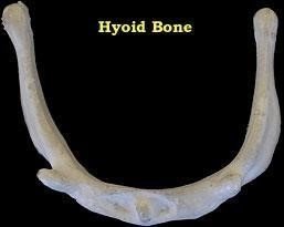
The Hyoid Bone
The hyoid bone is a unique U-shaped bone located in the anterior neck, inferior to the mandible. It does not articulate with any other bone and serves as an attachment site for muscles involved in swallowing and speech.
Supports the tongue.
Elevates the larynx during swallowing.
Indicator of strangulation if fractured.
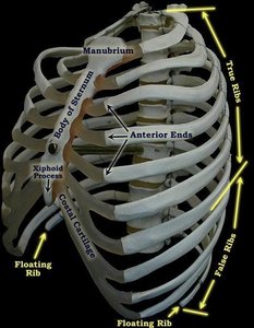
The Ribcage
The ribcage consists of the sternum and ribs, providing protection for the thoracic organs.
True ribs (1-7): Attach directly to the sternum via costal cartilage.
False ribs (8-12): Attach indirectly or not at all to the sternum.
Floating ribs (11-12): Do not attach to the sternum.
Costal cartilage: Connects ribs to the sternum.
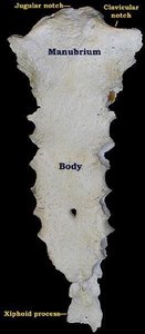
Sternum
The sternum is the breastbone, composed of three fused bones:
Manubrium: Superior portion with jugular and clavicular notches.
Body: Middle portion.
Xiphoid process: Inferior portion, important for CPR and Heimlich maneuver.
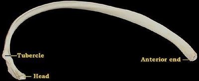
Rib Anatomy
Each rib has distinct features:
Anterior end: Articulates with costal cartilage.
Head: Articulates with thoracic vertebrae.
Tubercle: Articulates with the vertebra below the head.
Vertebral Column
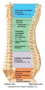
The vertebral column consists of 33 vertebrae grouped into regions:
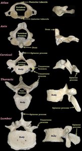
Cervical (7): Smallest, with transverse foramina; atlas (C1) and axis (C2) are specialized.
Thoracic (12): Larger, heart-shaped bodies, long spinous processes.
Lumbar (5): Largest, thick bodies, short spinous processes.
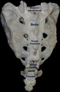
Sacrum (5 fused): Triangular, forms posterior pelvic wall.
Coccyx (4-5 fused): Tailbone, small and vestigial.
Bone Classification by Shape
Bones are classified by their shape, which relates to their function.
Long bones: Longer than wide; found in limbs (e.g., femur, humerus).
Short bones: Cube-shaped; found in wrists and ankles (e.g., carpals, tarsals).
Sesamoid bones: Form in tendons; patella is an example.
Flat bones: Thin, flattened; found in skull, sternum, ribs, scapulae.
Irregular bones: Complex shapes; vertebrae and hip bones.
Osseous Tissue (Bone Tissue)
Structure and Function
Bone tissue is composed of three cell types:
Osteocytes: Mature bone cells found in lacunae.
Osteoblasts: Bone-forming cells.
Osteoclasts: Bone-resorbing cells.
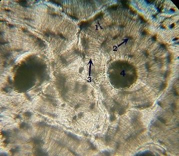
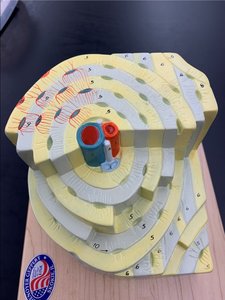
The basic unit of compact bone is the osteon (Haversian system), which consists of:
Central (Haversian) canal: Contains blood vessels and nerves.
Lamellae: Concentric rings of bony matrix.
Lacunae: Spaces housing osteocytes.
Canaliculi: Tiny canals connecting lacunae.
Appendicular Skeleton
Anatomy of a Long Bone
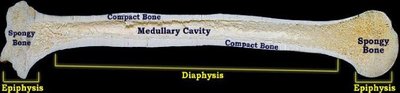
Long bones have a shaft (diaphysis) and two ends (epiphyses). The medullary cavity runs through the diaphysis, surrounded by spongy and compact bone.
Diaphysis: Shaft, provides leverage and weight support.
Epiphysis: Ends, contain spongy bone.
Medullary cavity: Contains bone marrow.
Shoulder Girdle
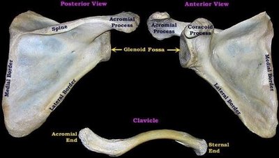
The shoulder girdle consists of the scapula and clavicle, providing attachment for upper limb muscles.
Scapula: Spine, acromial process, coracoid process, glenoid fossa, borders.
Clavicle: Acromial end (lateral), sternal end (medial).
Proximal Bones of the Limbs
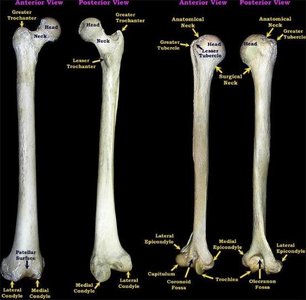
Femur: Head, neck, greater and lesser trochanters, condyles, patellar surface.
Humerus: Head, anatomical and surgical necks, tubercles, epicondyles, fossa, trochlea, capitulum.
Distal Bones of the Limbs
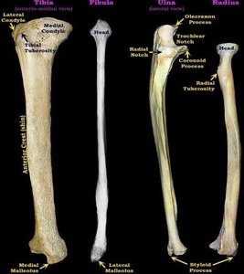
Tibia: Condyles, tuberosity, crest, malleolus.
Fibula: Head, lateral malleolus.
Ulna: Olecranon process, trochlear notch, coronoid process, radial notch, styloid process.
Radius: Head, radial tuberosity, styloid process.
The Pelvis
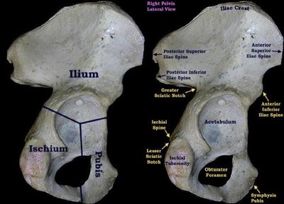
The pelvis is composed of the ilium, ischium, and pubis, which are fused.
Ilium: Iliac crest, spines.
Ischium: Ischial spine, tuberosity, sciatic notches.
Pubis: Symphysis pubis, obturator foramen.
Acetabulum: Socket for femur.
Male vs. Female Pelvis
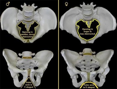
Male pelvis: Smaller, more obstructed opening, pubic arch is V-shaped.
Female pelvis: Larger, less obstructed opening, pubic arch is U-shaped.
Hands and Feet
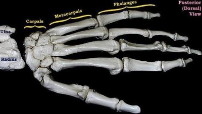
Hand: Carpals (wrist), metacarpals (palm), phalanges (fingers).
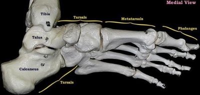
Foot: Tarsals (ankle), metatarsals (arch), phalanges (toes).
Articulations (Joints)
Synovial Joints
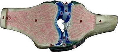
Synovial joints are freely movable and characterized by the presence of a synovial cavity.
Synovial fluid: Lubricates the joint.
Synovial membrane: Lines the joint capsule.
Meniscus: Cartilage pads for shock absorption.
Ligaments: Stabilize the joint.
Summary Table: Bone Shapes and Examples
Bone Shape | Description | Examples |
|---|---|---|
Long | Longer than wide | Femur, humerus |
Short | Cube-shaped | Carpals, tarsals |
Flat | Thin, flattened | Sternum, skull bones |
Irregular | Complex shape | Vertebrae, hip bones |
Sesamoid | Form in tendons | Patella |
Summary Table: Skull Sutures
Suture | Location |
|---|---|
Coronal | Between frontal and parietal bones |
Sagittal | Between parietal bones |
Lambdoid | Between parietal and occipital bones |
Squamous | Between parietal and temporal bones |
Summary Table: Vertebral Column Regions
Region | Number of Vertebrae | Key Features |
|---|---|---|
Cervical | 7 | Transverse foramina, bifid spinous process |
Thoracic | 12 | Heart-shaped body, long spinous process |
Lumbar | 5 | Large body, short spinous process |
Sacrum | 5 (fused) | Triangular, sacral foramina |
Coccyx | 4-5 (fused) | Small, vestigial |
Summary Table: Osseous Tissue Features
Feature | Description |
|---|---|
Osteocyte | Mature bone cell |
Lacuna | Space housing osteocyte |
Canaliculi | Tiny canals connecting lacunae |
Central canal | Contains blood vessels and nerves |
Lamellae | Concentric rings of matrix |
Key Equations and Concepts
Bone Density:
Osteon Structure:
Conclusion
The axial and appendicular skeletons provide structural support, protection, and facilitate movement. Understanding bone markings, shapes, and osseous tissue is essential for identifying bones and their functions in the human body. Additional info: Academic context and expanded explanations were added to ensure completeness and clarity for exam preparation.
