 뒤로
Axial and Appendicular Skeleton: Structure and Function
스터디 가이드 - 스마트 노트
 자료에 맞춘 맞춤형 노트, 핵심 정의, 예시, 맥락을 확장해 제공합니다.
The Human Skeleton: Overview
Axial and Appendicular Skeleton
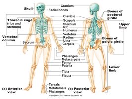
The human skeleton consists of 206 bones, accounting for about 20% of body weight. It is divided into two main regions: the axial skeleton and the appendicular skeleton. The axial skeleton includes the skull, vertebral column, and rib cage, while the appendicular skeleton comprises the bones of the limbs and the girdles that attach them to the axial skeleton.
Axial skeleton: Skull, vertebrae, rib cage
Appendicular skeleton: Upper and lower limbs, pectoral and pelvic girdles
Skull: Structure and Regions
Major Bones of the Skull
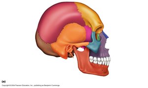
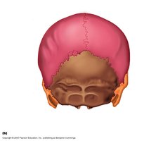
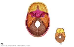
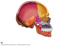
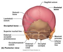
The skull is composed of several bones that are fused together. Different colored regions in anatomical diagrams correspond to distinct bones, such as the frontal, parietal, occipital, temporal, sphenoid, and ethmoid bones. These bones protect the brain and support facial structures.
Frontal bone: Forehead region
Parietal bones: Sides and roof of the cranium
Occipital bone: Posterior and base of the skull
Temporal bones: Sides of the skull, house ear structures
Sphenoid bone: Central, bat-shaped bone
Ethmoid bone: Deep, cube-shaped bone between orbits and nasal cavity
Temporal Bone
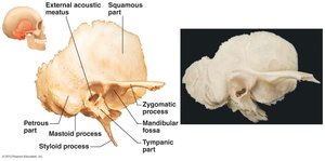
The temporal bone is irregularly shaped and serves as the site of attachment for neck and tongue muscles. It houses the middle and inner ear cavities, which are located in the petrous part—the hardest bone in the body, providing secure protection for organs involved in hearing and balance.
Sphenoid Bone
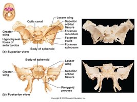
The sphenoid bone is a complex, bat-shaped bone that articulates with all other cranial bones. It forms part of the floor of the cranial cavity and contains important foramina for nerves and blood vessels.
Greater and lesser wings: Contribute to the cranial floor
Optic canal: Passage for optic nerve
Ethmoid Bone
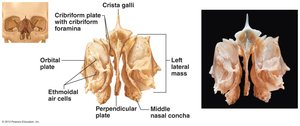
The ethmoid bone is cube-shaped and lies deep between the orbits and nasal cavities. The crista galli projects superiorly to attach to the dura mater of the brain. It contains the cribriform plate, which allows passage of olfactory nerves.
Sutural Bones
Sutural bones are tiny, irregular bones that can occur within cranial sutures. They result from additional ossification centers appearing rapidly during fetal development.
Hyoid Bone
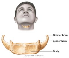
The hyoid bone is unique as it does not articulate directly with any other bone. It is anchored by ligaments and acts as a movable base for the tongue and a site of attachment for muscles involved in swallowing and speech. Its horseshoe shape makes it susceptible to fracture during strangulation.
Paranasal Sinuses
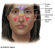
Paranasal sinuses lighten the skull, moisturize and warm incoming air, and contribute to voice resonance. The main sinuses are the frontal, ethmoidal, sphenoidal, and maxillary sinuses.
Vertebral Column
Structure and Regions
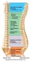
The vertebral column, or spine, extends from the skull to the pelvis. It transmits the weight of the trunk to the lower limbs, surrounds and protects the spinal cord, and provides attachment points for ribs and muscles. It consists of 33 bones, of which 24 remain separate for flexibility, while the remaining 9 fuse to form the sacrum and coccyx.
Cervical region: 7 vertebrae
Thoracic region: 12 vertebrae
Lumbar region: 5 vertebrae
Sacrum: 5 fused vertebrae
Coccyx: 3-5 fused vertebrae
Homeostasis and Vertebral Column
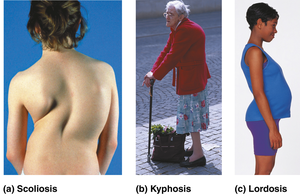
The vertebral column plays a crucial role in maintaining homeostasis by protecting the spinal cord and supporting the body's structure. Disorders such as scoliosis, kyphosis, and lordosis can affect posture and health.
Intervertebral Discs and Ligaments
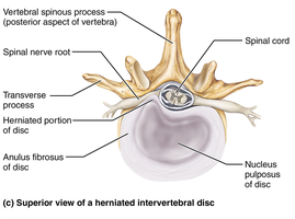
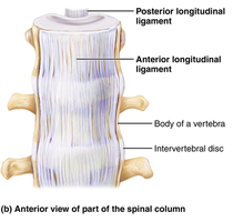
Intervertebral discs are cushion-like pads between vertebrae that act as shock absorbers. Ligaments support the column of bones, keeping it upright. The major supporting ligaments are the anterior and posterior longitudinal ligaments, which run along the front and back of the vertebral bodies.
Anterior longitudinal ligament: Broad, strongly attached; prevents hyperextension
Posterior longitudinal ligament: Narrow, weaker; prevents hyperflexion
General Structure of Vertebrae
All vertebrae share common features:
Body: Anterior, weight-bearing region
Vertebral arch: Composed of pedicles and laminae
Vertebral foramen: Enclosure formed by body and arch
Vertebral canal: Series of vertebral foramina
Intervertebral foramina: Lateral openings for spinal nerves
*Additional info: Regional differences exist among cervical, thoracic, and lumbar vertebrae, affecting movement and function.*
Sacrum and Coccyx
The sacrum starts as five separate vertebrae that fuse during adolescence. It serves as a central support for the skeleton and houses nerves. The coccyx, or tailbone, is formed from three to five fused vertebrae and provides support while sitting and serves as an anchor for pelvic floor muscles.
*Additional info: The sacrum articulates with the pelvic girdle, forming the posterior wall of the pelvis.*
Thoracic Cage: Sternum and Ribs
Structure and Function
The thoracic cage is composed of thoracic vertebrae, ribs, sternum, and costal cartilages. It protects vital organs, supports the shoulders and upper limbs, and provides attachment sites for muscles.
Sternum: Breastbone, consists of manubrium, body, and xiphoid process
Ribs: Shaft, head, neck, tubercle; articulate with vertebrae
*Additional info: During CPR, hands must be placed on the midsternum to avoid breaking the xiphoid process.*
Appendicular Skeleton
Pectoral and Pelvic Girdles
The appendicular skeleton includes the bones of the limbs and their girdles. The pectoral girdle attaches the upper limbs to the trunk, while the pelvic girdle attaches the lower limbs. The pelvic girdle is formed by two hip bones and the sacrum, providing stability and support for pelvic organs.
*Additional info: The pelvic girdle transmits weight from the upper body to the lower limbs and is less mobile but more stable than the shoulder joint.*
Bones of the Upper Limb
The upper limb consists of 30 bones:
Arm: Humerus
Forearm: Radius and ulna
Hand: Carpals (wrist), metacarpals (palm), phalanges (fingers)
*Additional info: The humerus is the longest bone in the upper limb; radius and ulna are parallel long bones.*
Bones of the Lower Limb
The lower limb carries the entire weight of the body and is subjected to exceptional forces. It consists of three segments: thigh (femur), leg (tibia and fibula), and foot (tarsals, metatarsals, phalanges).
Tibia: Medial, weight-bearing bone
Fibula: Lateral, not weight-bearing
*Additional info: The patella is the kneecap; the foot includes tarsals, metatarsals, and phalanges.*
Pelvis: True vs. False Pelvis and Sex Differences
The pelvis is divided into the true and false pelvis. Structural differences exist between male and female pelves, affecting childbirth and locomotion.
*Additional info: The true pelvis is the region below the pelvic brim; the false pelvis is above it.*
Summary
This lecture covered the anatomy of the axial and appendicular skeleton, focusing on the structure and function of bones, their regions, and their physiological relevance to human health and homeostasis.
